. 2021 Jan 25;49(2):805-817.
doi: 10.1093/nar/gkaa1243.

The fission yeast Pin1 peptidyl-prolyl isomerase promotes dissociation of Sty1 MAPK from RNA polymerase II and recruits Ssu72 phosphatase to facilitate oxidative stress induced transcription

Affiliations

The fission yeast Pin1 peptidyl-prolyl isomerase promotes dissociation of Sty1 MAPK from RNA polymerase II and recruits Ssu72 phosphatase to facilitate oxidative stress induced transcription

Yi-Ting Wang et al. Nucleic Acids Res. .

Abstract

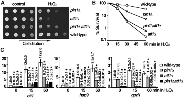
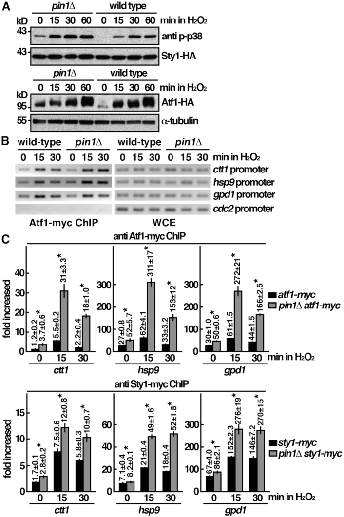
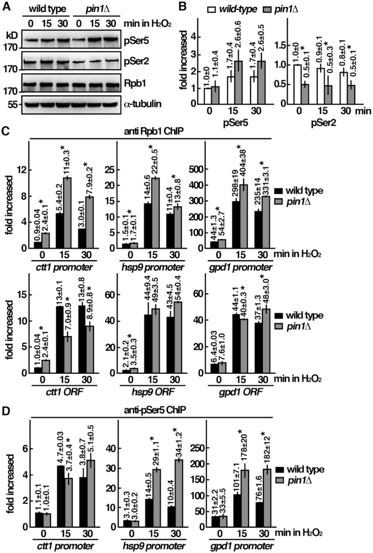
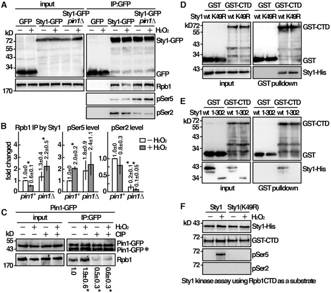
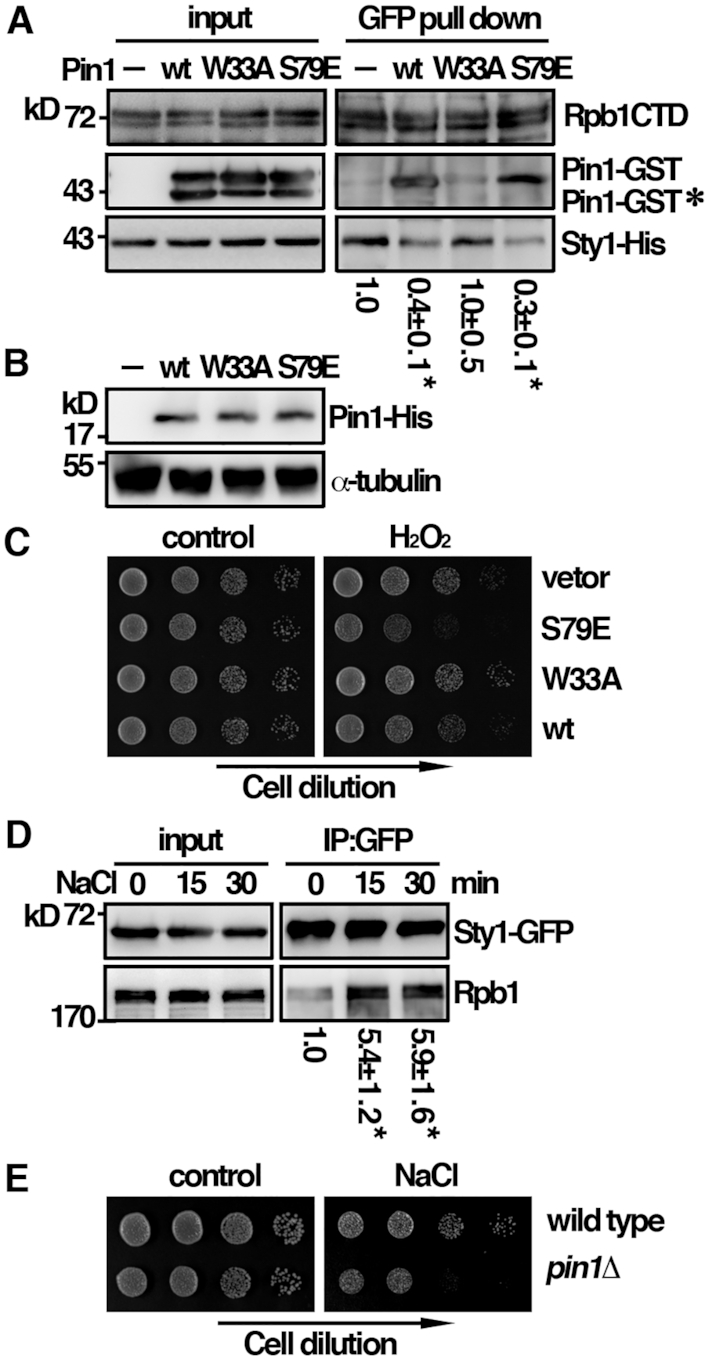
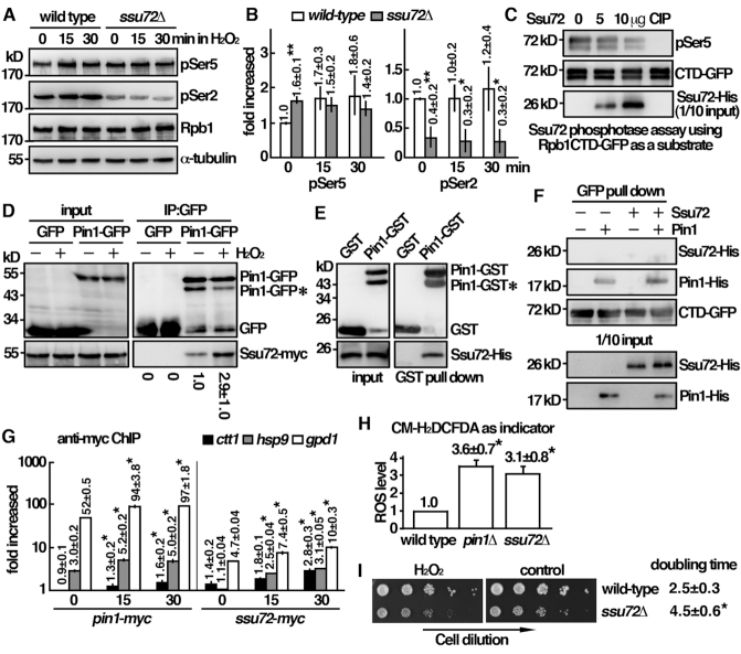
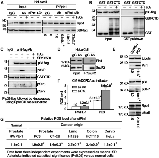
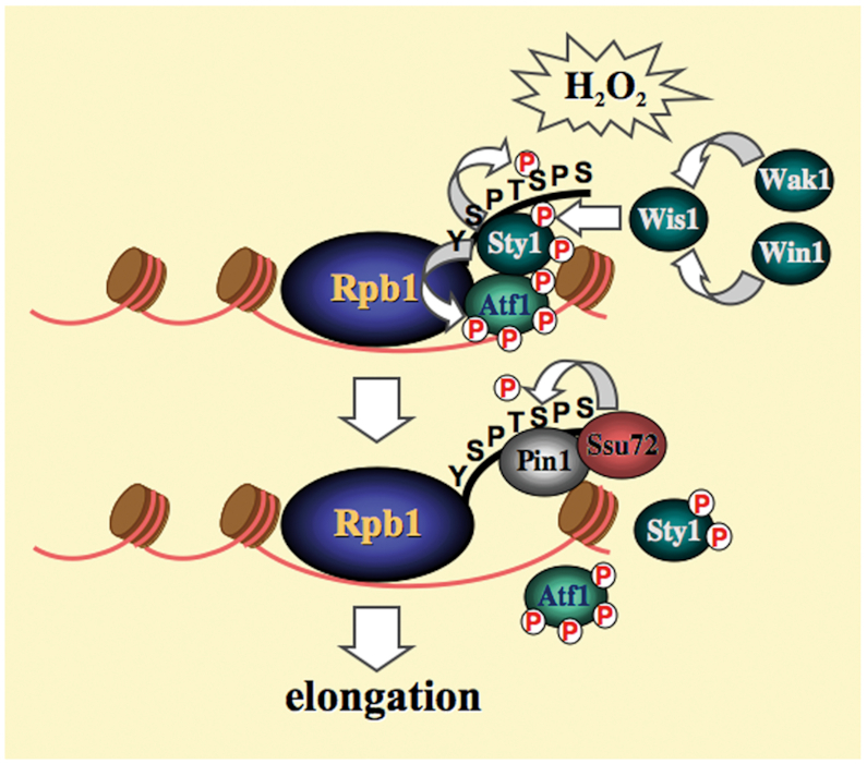
Pin1 is a peptidyl-prolyl isomerase that regulates the structure and function of eukaryotic RNA polymerase II (Pol II) through interaction with the C-terminal domain (CTD) of Rpb1, the largest subunit of Pol II. We demonstrated that this function is important for cellular response to oxidative stress in the fission yeast Schizosaccharomyces pombe. In response to oxidative stress, the Atf1 transcription factor targets Sty1, the mitogen-activated protein kinase (MAPK), to specific stress-responsive promoters. Anchored Sty1 recruits Pol II through direct association with Rpb1-CTD and phosphorylates the reiterated heptad sequence at Serine 5. Pin1 binds phosphorylated CTD to promote dissociation of Sty1 from it, and directly recruits Ssu72 phosphatase to facilitate dephosphorylation of CTD for transcription elongation. In the absence of Pin1, the association of Sty1-Atf1 with Rpb1 persists on stress-responsive promoters failed to generate transcripts of the corresponding genes effectively. The identified characteristic features of the fission yeast Pin1 are conserved in humans. We demonstrated that elevated Pin1 level in cancer cells might help to sustain survival under oxidative stress generated from their altered metabolic pathways. Together, these results suggest a conserved function of Pin1 in cellular response to oxidative stress among eukaryotic cells that might have clinical implication.


Figures

Figure 1.
Figure 1.
Pin1 mutant is defective in oxidative stress induced transcription. (A) Tenfold serial dilution of strains indicated were spotted onto YES-agar plates without (control) or with 2 mM H2O2 and photographed after 3 days incubation at 30°C. (B) H2O2 (2 mM) was added to an asynchronous culture of strains indicated growing at 30°C in YES medium. Cell viability was measured at the time indicated by plating appropriate dilution of cells onto YES agar plate without drug and scoring colony formation after 3 days incubation at 30°C. Viability (% survival) is expressed as a percentage of the number of colonies obtained before the addition of drug. (C) Expression of stress-responsive genes indicated upon oxidative stress in wild type, pin1Δ, atf1Δ and pin1Δatf1Δ cells. Total RNA from cultures of the indicated strains grown at 30°C was reverse transcribed using an oligo(dT) primer. The cDNA was amplified using quantitative PCR and SYBR green with primers specific for the indicated genes. The amount of RNA normalized to cdc2 mRNA relative to the wild type was expressed as means ± SD in triplicate. Asterisks indicated statistical significance (P< 0.05, t-test unpaired) verse wild type.
Figure 2.
Figure 2.
Pin1 mutant is not defective in MAPK-mediated signal transduction pathway. (A) Whole-cell protein extracts of the strains indicated were prepared by alkaline extraction followed by trichloroacetic acid precipitation. The extracts were separated by SDS-PAGE and subjected to Western immunoblotting using anti-HA antibodies to reveal proteins of interest. Anti p-p38 antibody cross-reacted with phosphorylated Sty1. Antibody against α-tubulin was used as the loading control. (B) ChIP assays showing recruitment of Atf1-myc to the promoter of ctt1, gpd1, hsp9 and cdc2 genes upon oxdative stress. The latter was not induced upon stress. Samples were prepared from atf1-myc and atf1-myc pin1Δ cells treated with 2 mM H2O2 for the time points indicated. The DNA recovered from the IP was assayed by PCR using primers specific to the promoter of genes indicated. The control lanes show DNA amplified from whole cell extracts (WCE) prior to performing the IP. (C) ChIP assays showing recruitment of Atf1-myc and Sty1-myc to the promoter of ctt1, gpd1 and hsp9 genes upon oxdative stress in pin1Δ mutant and wild type cells. Samples were prepared from strains indicated treated with 2 mM H2O2 for the time points indicated. The DNA recovered from the IP was amplified using quantitative PCR and SYBR green with primers specific to the promoter of genes indicated normalized to that of the non-stress responsive cdc2 gene. Data were expressed as means ± SD in triplicate. Asterisks indicated statistical significance (P< 0.05, t-test unpaired) verse wild type.
Figure 3.
Figure 3.
Pin1 mutant is defective in the oxidative stress induced transcription response at the initiation to elongation transition. (A) Whole-cell protein extracts of the strains indicated were prepared by alkaline extraction followed by trichloroacetic acid precipitation. The extracts were separated by SDS-PAGE analyzed phosphorylation by western immunoblotting with CTD phospho-specific antibodies. Antibody against α-tubulin was used as the control. The relative level of phosphorylated protein was shown in (B). Data were expressed as means±SD in triplicate. Asterisks indicated statistical significance (P<0.05, t-test unpaired) verse wild type. (C) ChIP assays showing the recruitment of Rpb1 to the promoter and the gene coding regions (ORF) of ctt1, gpd1 and hsp9 genes upon oxidative stress in wild type cells and pin1Δ mutant. Samples were prepared from the indicated strains treated with 2 mM H2O2 for the time points indicated. The DNA recovered from the IP was amplified using quantitative PCR and SYBR green with primers specific to the promoter of the indicated genes normalized to that of the non-stress responsive cdc2 gene. Data were expressed as means ± SD in triplicate. Asterisks indicated statistical significance (P< 0.05, t-test unpaired) verse wild type. (D) ChIP assays showing the recruitment of pSer5-Rpb1 to the promoters of ctt1, gpd1 and hsp9 genes upon oxdative stress in wild type cells and pin1Δ mutant as in (C). Data were expressed as means ± SD in triplicate. Asterisks indicated statistical significance (P< 0.05, t-test unpaired) verse wild type.
Figure 4.
Figure 4.
Sty1 interacted and phosphorylated Rpb1-CTD at Ser5. (A) Coimmunoprecipitation was performed with extracts prepared from indicated strains treated with 2 mM H2O2 for 30 min. GFP-Trap affinity resin was used to pull down GFP proteins. Immunoprecipitates were analyzed by Western immunoblotting using antibodies against GFP and Rpb1-CTD. The relative level of Rpb1 pull down by Sty1 and its phosphorylation status was shown in (B). Data were expressed as means±SD in triplicate. Asterisks indicated statistical significance (P< 0.05, t-test unpaired) verse wild type. (C) Coimmunoprecipitation was performed with extracts prepared from Pin1-GFP tagged strains treated with 2 mM H2O2 for 30 min. GFP-Trap affinity resin was used to pull down GFP proteins. Immunoprecipitates were treated with/without CIP and analyzed by Western immunoblotting using antibodies against GFP and Rpb1-CTD. *Degradation product of Pin1-GFP. The relative level of Rpb1 pull down by Pin1 was shown. Data were expressed as means±SD in triplicate. Asterisks indicated statistical significance (P<0.05, t-test unpaired) verse control of no treatments. (D, E) Purified GST-CTD from Escherichia coli and recombinant protein of Sty1 from yeast cells were subjected to GST pulldown assay followed by Western immunoblotting analysis with antibodies against GST and His. (F) In vitro kinase assay using Sty1-His extracted and immunoprecipitated from activated cells. Following incubation with GST-CTD, proteins were resolved by SDS-PAGE and probed with antibodies indicated.
Figure 5.
Figure 5.
A mutual exclusive binding mode of Rpb1 to Pin1 over Sty1. (A) Recombinant proteins of Sty1 and CTD-GFP from yeast cells were subjected to GFP pulldown assay in the presence or absence of Pin1 followed by Western immunoblotting analysis with antibodies indicated. *Degradation product of Pin1-GST. The relative level of Sty1 pull down by CTD-GFP was shown. Data were expressed as means±SD in triplicate. Asterisks indicated statistical significance (P<0.05, t-test unpaired) verse control without Pin1. (B) Whole-cell protein extracts of the strains indicated were prepared by alkaline extraction followed by trichloroacetic acid precipitation. The extracts were separated by SDS-PAGE and subjected to immunoblotting using anti-His antibodies to reveal Pin1 proteins. Antibody against α-tubulin was used as the control. (C) Tenfold serial dilutions of strains indicated were spotted onto EMM+thiamine agar plates without (control) or with 2 mM H2O2 and photographed after 3 days incubation at 30°C. (D) Coimmunoprecipitation was performed with extracts prepared from Sty1-GFP tagged strains treated with 0.5 M NaCl for the time point indicated. GFP-Trap affinity resin was used to pull down GFP proteins. Immunoprecipitates were analyzed by Western immunoblotting using antibodies against GFP and Rpb1-CTD. The relative level of Rpb1 pull down by Sty1 was shown. Data were expressed as means±SD in triplicate. Asterisks indicated statistical significance (P< 0.05, t-test unpaired) verse control of no treatment. (E) Tenfold serial dilutions of strains indicated were spotted onto YES-agar plates without (control) or with 200 mM NaCl and photographed after 3 days incubation at 30°C.
Figure 6.
Figure 6.
Pin1 directly interacts and recruits Ssu72 for pSer5 dephosphorylation. (A) Whole-cell protein extracts of the strains indicated were prepared by alkaline extraction followed with trichloroacetic acid precipitation. The extracts were separated by SDS-PAGE and analyzed phosphorylation by Western immunoblotting with CTD phospho-specific antibodies. Antibody against α-tubulin was used as the control. The relative level of phosphorylated protein was shown in (B). Data were expressed as means±SD in triplicate. Asterisks indicated statistical significance (P< 0.05, t-test unpaired) verse wild type. (C) In vitro phosphatase activity assay using recombinant protein of CTD-GFP from yeast cells were treated with Ssu72 recombinant protein or 20 units calf intestinal alkaline phosphatase (CIP) at 30°C for 1 h. Reaction mixtures were resolved by SDS-PAGE and probed with antibodies indicated. (D) Coimmunoprecipitation was performed with extracts prepared from Ssu72-myc tagged strains expressing Pin1-GFP and being treated with 2 mM H2O2 for 30 min. GFP-Trap affinity resin was used to pull down GFP proteins. Immunoprecipitates were analyzed by western immunoblotting using antibodies against GFP and myc. *Degradation product of Pin1-GFP. The relative level of Ssu72 pull down by Pin1-GFP was shown. Data were expressed as means±SD in triplicate. (E) Recombinant proteins of Pin1-GST and Ssu72-His were subjected to GST pull down assay followed by Western immunoblotting analysis with antibodies against GST and His. *Degradation product of Pin1-GST. (F) Recombinant proteins of Ssu72-His and Pin1-His were subjected to GFP pulldown assay by CTD-GFP followed by Western immunoblotting analysis with antibodies against GFP and His. (G) ChIP assays showing the recruitment of Pin1-myc and Ssu72-myc to the promoters of ctt1, gpd1 and hsp9 genes upon oxdative stress. Samples were prepared from strains indicated treated with 2 mM H2O2 for the time points indicated. The DNA recovered from the IP was amplified using quantitative PCR and SYBR green with primers specific to the promoters of genes indicated normalized to that of the non-stress responsive cdc2 gene. Data was expressed as means ± SD in triplicate. Asterisks indicated statistical significance (P< 0.05, t-test unpaired) verse control of no treatment. (H) Strains indicated were subjected to measurement of intracellular ROS levels in triplicate using CM-H2DCFDA as indicator. Asterisks indicated statistical significance (P< 0.05, t-test unpaired) verse wild type. (I) Tenfold serial dilutions of strains indicated were spotted onto YES-agar plates without (control) or with 2 mM H2O2 and photographed after 3 days incubation at 30°C. Growth characteristics of the strains indicated at 30°C (doubling time) were shown on the right. Data were expressed as means ± SD in triplicate. Asterisks indicated statistical significance (P< 0.05, t-test unpaired) verse wild type.
Figure 7.
Figure 7.
The characteristic feature of the fission yeast Pin1 identified is conserved in humans. (A) Coimmunoprecipitation was performed with extracts prepared from human HeLa cells expressing p38 tagged with flag. Immunoprecipitates were analyzed by Western immunoblotting analysis using antibodies specific for Rpb1 and flag. The relative level of Rpb1 pull down by p38 was shown. Data were expressed as means±SD in triplicate. Asterisks indicated statistical significance (P< 0.05, t-test unpaired) verse control of no treatment. (B) Purified GST-CTD from Escherichia coli and p38-flag precipitated from HeLa cells were subjected to GST pulldown assay followed by Western immunoblotting analysis with antibodies against GST and flag. (C) In vitro kinase assay using p38 extracted and immunoprecipitated from activated cells. Following incubation with GST-CTD, proteins were resolved by SDS-PAGE and probed with antibodies indicated. (D) Coimmunoprecipitation was performed with extracts prepared from human HeLa cells using anti-Ssu72 antibody and IgG as control. Immunoprecipitates were analyzed by Western immunoblotting analysis with antibodies specific for human Pin1 and Ssu72. (E) Total lysates of human cell indicated were subjected to Western immunoblotting analysis with indicated antibodies. The relative level of protein of interest was indicated beneath each lane. Data were expressed as means±SD in triplicate. Asterisks indicated statistical significance (P< 0.05, t-test unpaired) verse RWPE-1 cells. (F) (G) Human cells indicated were subjected to measurement of intracellular ROS Levels in triplicate using CM-H2DCFDA as indicator. Data were expressed as means±SD in triplicate. Asterisks indicated statistical significance (P< 0.05, t-test unpaired) verse normal RWPE-1 cells.
Figure 8.
Figure 8.
Model of Sty1-mediated oxidative stress induced transcription. Upon oxidative stress, activated Sty1 phosphorylates the CTD reiterated heptad sequence at Serine 5. Pin1 competes the binding of phosphorylated CTD to promote Sty1 dissociating from it, and directly recruits Ssu72 to facilitate dephosphorylation of CTD for transcription elongation.

Similar articles

Cited by

References

    1. Derijard B., Hibi M., Wu I.H., Barrett T., Su B., Deng T., Karin M., Davis R.J.. JNK1: a protein kinase stimulated by UV light and Ha-Ras that binds and phosphorylates the c-Jun activation domain. Cell. 1994; 76:1025–1037. - PubMed
    1. Livingstone C., Patel G., Jones N.. ATF-2 contains a phosphorylation-dependent transcriptional activation domain. EMBO J. 1995; 14:1785–1797. - PMC - PubMed
    1. Shiozaki K., Russell P.. Cell-cycle control linked to extracellular environment by MAP kinase pathway in fission yeast. Nature. 1995; 378:739–743. - PubMed
    1. Reiter W., Watt S., Dawson K., Lawrence C.L., Bahler J., Jones N., Wilkinson C.R.. Fission yeast MAP kinase Sty1 is recruited to stress-induced genes. J. Biol. Chem. 2008; 283:9945–9956. - PMC - PubMed
    1. Alepuz P.M., de Nadal E., Zapater M., Ammerer G., Posas F.. Osmostress-induced transcription by Hot1 depends on a Hog1-mediated recruitment of the RNA Pol II. EMBO J. 2003; 22:2433–2442. - PMC - PubMed

Publication types

MeSH terms