Case Reports
. 2021 Oct;479(4):827-833.
doi: 10.1007/s00428-020-02975-6. Epub 2021 Jan 7.

Transbronchial biopsies' histopathological findings leading to successful late steroid therapy in Covid-19 acute respiratory failure

Affiliations
Case Reports

Transbronchial biopsies' histopathological findings leading to successful late steroid therapy in Covid-19 acute respiratory failure

Francisco Klein et al. Virchows Arch. 2021 Oct.

Abstract

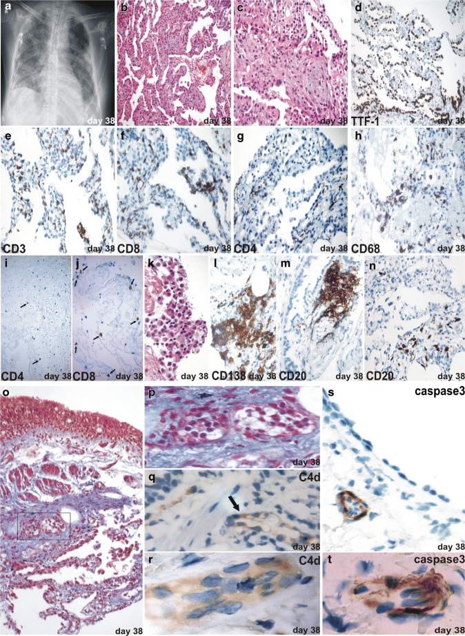
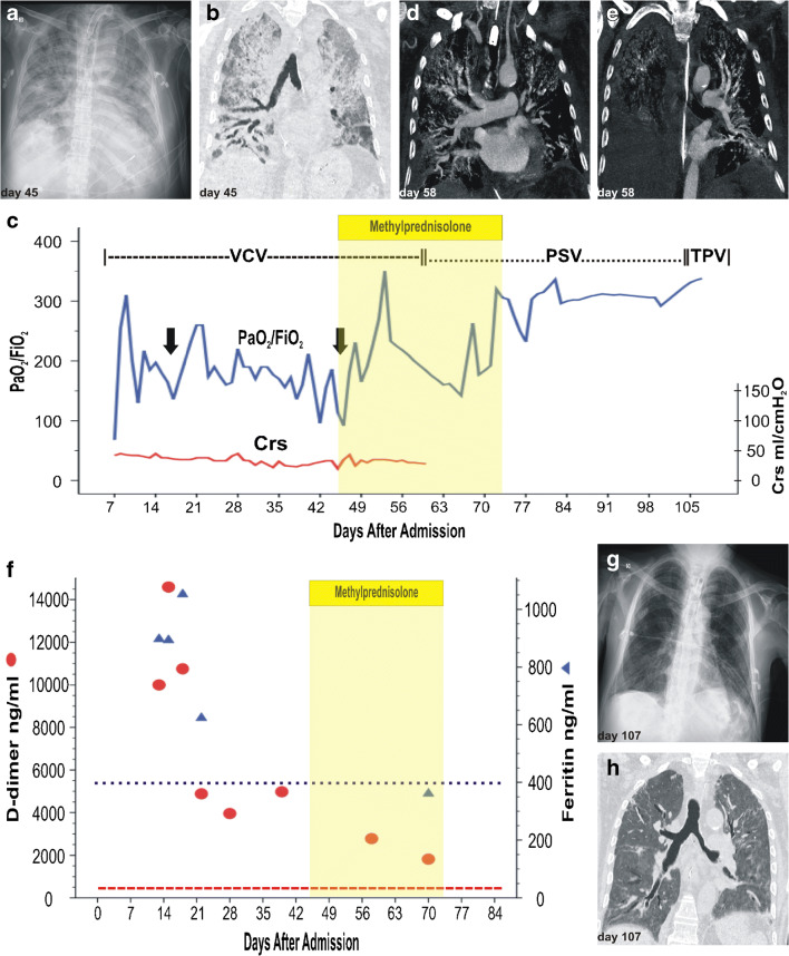
We present results from clinical, radiologic, gas exchange, lung mechanics, and fibre-optic bronchoscopy-guided transbronchial biopsies in a case of acute respiratory failure due to SARS-CoV-2 (Covid-19). This report highlights the pulmonary, immunological, and inflammatory changes found during acute diffuse alveolar damage and the later organizing phase. An early diffuse alveolar damage pattern with predominant epithelial involvement with active recruitment of T cells and monocytes was observed followed by a late organizing pattern with pneumocyte hyperplasia, inflammatory infiltration, prominent endotheliitis, and secondary germinal centers. The patient's deterioration paralleling the late immuno-pathological findings based the decision to administer intravenous corticosteroids, resulting in clinical, gasometric, and radiologic improvement. We believe that real-time clinicopathological correlation, along with the description of the immunological processes at play, will contribute to the full clinical picture of Covid-19 and might lead to a more rational approach in the precise timing of anti-inflammatory, anti-cytokine, or steroid therapies.

Keywords: Acute respiratory failure; Covid-19; Lung histology; Pathology; SARS-CoV-2; Transbronchial biopsy.


Conflict of interest statement

The authors declare that they have no conflict of interest.

Figures

Fig. 1
Fig. 1
Early phase. a Hospital day 3 CXR. Right basilar opacity. b Hospital day 3 chest CT. Bilateral and peripheral ground-glass opacities, mainly in the right lower lobe. c Hospital day 7 CXR. Infiltrates of diffuse bilateral distribution with consolidation associated with air bronchogram at the right perihilar level. d Hospital day 7 chest CT. Increasing bilateral ground-glass opacities with predominant subpleural distribution; thickening of the inter and intralobular septa, showing a “crazy paving” pattern. Air space consolidation, air bronchogram, and thickening of the bronchial walls. e Timeline chart of white-cell count in peripheral blood (hospital days 1 to 7). f BAL, CD4+ T cells (arrows), immunohistochemical stain (IHC) (× 400). g BAL CD8+ T cells (arrows), IHC (× 400). h Lung parenchyma with detachment of pneumocytes and formation of hyaline membranes, moderate mono and polymorphonuclear infiltrate, consistent with an early phase of diffuse alveolar damage. H&E (× 200). Insert, pneumocytes with enlarged nuclei showing cytopathic changes from a likely viral origin (arrows). i Bronchial wall, lamina propria, infiltrate of CD3+ T cells, IHC (× 400). j Bronchial wall, epithelium, and lamina propria, infiltrate of CD8+ T cells, IHC (× 400). k Bronchial wall, lamina propria, macrophage infiltrate (CD68+), IHC (× 400). l Bronchial wall, lamina propria, infiltrate of CD4+ T cells, IHC (× 400). m Lung parenchyma, infiltrate of CD3+ T cells, IHC (× 400). n Lung parenchyma, infiltrate of CD8+ T cells, IHC (× 400)
Fig. 2
Fig. 2
Late phase. a CXR. Evidence of increased infiltrates, lung consolidation, and bibasal bronchial dilation. b Diffuse thickening of the interalveolar septa with fibromyxoid material and mononuclear inflammatory infiltrate. H&E (× 200) and c H&E (× 400). d Lung parenchyma, type II pneumocyte hyperplasia, positive for TTF1, IHC (× 200). e Alveolar septa, infiltrate of CD3+ T cells, IHC (× 400). f Alveolar septa, infiltrate of CD8+ T cells, IHC (× 400). g Alveolar septa, infiltrate of CD4+ T cells, IHC (× 400). h Alveolar septa, infiltrate of CD68+ T cells, IHC (× 400). i BAL, CD4+ T cells (arrows), IHC (× 400). j BAL CD8+ T cells (arrows), IHC (× 400). k BAL with clusters of plasma cells, mixed with macrophages and lymphocytes, H&E (× 400). l Lung parenchyma with plasma cell clusters, positive for CD138, IHC (× 400). m Bronchial wall, CD20+ B cell aggregates in lamina propria, IHC (× 400). n Lung parenchyma, infiltrate of CD20+ B cells, IHC (× 400). o Lung parenchyma, with endotheliitis with predominantly intraluminal neutrophils (box). Masson trichrome (× 200) and p (× 1000). q, r C4d deposition is seen in vessels of the lung parenchyma (arrow). IHC (× 1000). s, t IHC staining caspase-3 in scattered lung vessels endothelial cells. HC (× 400) and (× 1000)
Fig. 3
Fig. 3
Follow-up and outcome. a Hospital day 45 CXR. Bilateral interstitial-alveolar infiltrates with areas of consolidation with air bronchogram. Obliteration of left costophrenic sinus and bibasilar bronchial dilation. b Hospital day 45 chest CT. Extensive progressive infiltrates with “crazy paving” pattern associated with consolidations. Bilateral pleural effusion associated with areas of passive atelectasis. Bronchial dilations and subpleural cysts are also observed, predominantly in the middle and lower right lobe. c PaO2/FiO2 ratio and total respiratory system compliance (Crs) curves (hospital days 7 to 107). On days 17 and 46 (arrows), 16/24-h prone positioning was indicated due to refractory hypoxaemia. Administration of 2 mg/kg/day of intravenous methylprednisolone (days 45–73). VCV, volume-controlled ventilation; PSV, pressure support ventilation; TPV, T piece ventilation. d, e Hospital day 58 HRCT angiography ruled out acute pulmonary embolism. f D-dimer and ferritin levels decreased toward normal values after steroid administration (days 45–73). g Hospital day 107 CXR. Marked reduction of alveolar interstitial infiltrates. h Hospital day 107 chest CT. Partial resolution of the diffuse inflammatory/infectious process visualized in previous examinations, persisting at the bibasal level. Thickening of the interlobular septa, associated with traction bronchiectasis and areas of honeycombing

References

    1. Calabrese F, Pezzuto F, Fortarezza F, Hofman P, Kern I, Panizo A, von der Thüsen J, Timofeev S, Gorkiewicz G, Lunardi F (2020) Pulmonary pathology and COVID-19: lessons from autopsy. The experience of European Pulmonary Pathologists. Virchows Arch 1–14. 10.1007/s00428-020-02886-6 - PMC - PubMed
    1. Tian S, Hu W, Niu L, Liu H, Xu H, Xiao SY. Pulmonary pathology of early-phase 2019 novel coronavirus (COVID-19) pneumonia in two patients with lung cancer. J Thorac Oncol. 2020;15(5):700–704. doi: 10.1016/j.jtho.2020.02.010. - DOI - PMC - PubMed
    1. Pernazza A, Mancini M, Rullo E, Bassi M, De Giacomo T, Rocca CD, d'Amati G (2020) Early histologic findings of pulmonary SARS-CoV-2 infection detected in a surgical specimen. Virchows Arch 1–6. 10.1007/s00428-020-02829-1 - PMC - PubMed
    1. Ministerio de Salud, Argentina (2020) nuevo-coronavirus-covid-19-reporte-diario. https://www.argentina.gob.ar/coronavirus/informe-diario. Accessed 26 July 2020
    1. Wang D, Hu B, Hu C, Zhu F, Liu X, Zhang J, Wang B, Xiang H, Cheng Z, Xiong Y, Zhao Y, Li Y, Wang X, Peng Z. Clinical characteristics of 138 hospitalized patients with 2019 novel coronavirus-infected pneumonia in Wuhan, China. JAMA. 2020;323(11):1061–1069. doi: 10.1001/jama.2020.1585. - DOI - PMC - PubMed

Publication types

MeSH terms

LinkOut - more resources