Review
. 2021 Jan 7;4(1):10.
doi: 10.1186/s42155-020-00176-5.

Renovascular hypertension in children

Affiliations
Review

Renovascular hypertension in children

Premal Amrishkumar Patel et al. CVIR Endovasc. .

Abstract

Paediatric hypertension, defined as systolic blood pressure > 95th percentile for age, sex and height is often incidentally diagnosed. Renovascular hypertension (RVH) is responsible for 5-25% of hypertension in children. Renal artery stenosis and middle aortic syndrome can both can be associated with various conditions such as fibromuscular dysplasia, Williams syndrome & Neurofibromatosis type 1. This paper discusses the approaches to diagnosis and interventional management and outcomes of renovascular hypertension in children. Angiography is considered the gold standard in establishing the diagnosis of renovascular disease in children. Angioplasty is beneficial in the majority of patients and generally repeated angioplasty is considered more appropriate than stenting. Surgical options should first be considered before placing a stent unless there is an emergent requirement. Given the established safety and success of endovascular intervention, at most institutions it remains the preferred treatment option.

Keywords: Fibro-muscular dysplasia; Mid-aortic syndrome; Paediatric interventional radiology; Renal artery stenosis.


Conflict of interest statement

Not applicable.

Figures

Fig. 1
Fig. 1
Renal Artery Stenosis Angioplasty. a Axial contrast enhanced CT angiogram image demonstrating a left renal artery stenosis with post stenotic dilation. b This was confirmed on subsequent rotational angiography and (c) at angioplasty by demonstration of a waist on the angioplasty balloon
Fig. 2
Fig. 2
Intravascular ultrasound. a IVUS image of severe right renal artery origin stenosis with mural thickening consistent with FMD. b Post angioplasty IVUS image of improved flow and vessel diameter right renal artery origin
Fig. 3
Fig. 3
Contrast enhanced ultrasound. Single frames from a contrast enhanced US video loops taken immediately after sulfur hexafluoride lipid-type A microspheres are injected and flushed with 10mls saline. a Immediately pre right renal artery angioplasty with reduced cortical perfusion to the right kidney and b immediately post angioplasty with overall improved perfusion
Fig. 4
Fig. 4
Angiographic perfusion imaging. Images of pre and post angioplasty angiography with post processing using Syngo I flow (Siemens AX) non parametric imaging data: time to peak opacification demonstrating using color coding and time points demonstrating improved time to renal perfusion post right renal accessory stenosis angioplasty
Fig. 5
Fig. 5
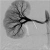
Non-flow limiting dissection. Angiography right renal artery demonstrating a lucent dissection flap with normal distal renal perfusion consistent with a non-flow limiting dissection (arrow)
Fig. 6
Fig. 6
Pseudoaneursym. a Left renal artery angiogram demonstrating early bifurcation with tight long segment stenosis of both renal artery branches (arrow). b Follow up 2 month angiogram demonstrates a pseudoaneurysm at the site of angioplasty of the upper renal artery branch, treated successfully with coil embolization (c)
Fig. 7
Fig. 7
Distal embolic phenomenon. Post right renal artery angioplasty demonstrating focal segmental renal artery occlusion (short arrow) with absent perfusion consistent with a distal renal thrombo-embolic event post angioplasty (long arrow)

Similar articles

Cited by

References

    1. Agrawal H, Moodie D, Qureshi AM, Acosta AA, Hernandez JA, Braun MC, Justino H (2018) Interventions in Children with Renovascular Hypertension: A 27-Year Retrospective Single-Center Experience. Congenit Heart Dis 13 (3):349–56. 10.1111/chd.12608 - DOI - PubMed
    1. Alexander A, Richmond L, Geary D, Salle JLP, Amaral J, Connolly B. Outcomes of percutaneous transluminal angioplasty for pediatric renovascular hypertension. J Pediatr Surg. 2017;52:395–399. doi: 10.1016/j.jpedsurg.2016.08.011. - DOI - PubMed
    1. Alhadad A, Ahle M, Ivancev K, Gottsäter A, Lindblad B. Percutaneous transluminal renal angioplasty (PTRA) and surgical revascularisation in renovascular disease--a retrospective comparison of results, complications, and mortality. Eur J Vasc Endovasc Surg Off J Eur Soc Vasc Surg. 2004;27:151–156. doi: 10.1016/j.ejvs.2003.10.009. - DOI - PubMed
    1. Arce-Santiago M, Rodríguez-Cruz E. Treatment of a recurrent renal artery stenosis and stent fracture using a drug eluting stent in a pediatric patient. CEN Case Rep. 2015;5:18–22. doi: 10.1007/s13730-015-0182-1. - DOI - PMC - PubMed
    1. Arend WP, Michel BA, Bloch DA, Hunder GG, Calabrese LH, Edworthy SM, et al. The American College of Rheumatology 1990 criteria for the classification of takayasu arteritis. Arthritis Rheum. 1990;33:1129–1134. doi: 10.1002/art.1780330811. - DOI - PubMed

LinkOut - more resources