

Case Reports
doi: 10.1212/NXI.0000000000000929.
Print 2021 Jan.
Treating MS after surviving PML: Discrete strategies for rescue, remission, and recovery patient 1: From the National Multiple Sclerosis Society Case Conference Proceedings
Affiliations
- PMID: 33411672
- PMCID: PMC7803340
- DOI: 10.1212/NXI.0000000000000929
Item in Clipboard
Case Reports
Treating MS after surviving PML: Discrete strategies for rescue, remission, and recovery patient 1: From the National Multiple Sclerosis Society Case Conference Proceedings
Neurol Neuroimmunol Neuroinflamm.
.
No abstract available
Figures
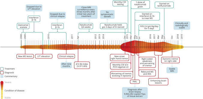
In this figure, we detail the condition of the patient over time. The
longitudinal axis (left to right) depicts the condition of disease,
where the smaller amplitude and lighter color indicates greater
stability of MS. Alternately, the expanded amplitude of the colored heat
map (above and below the horizontal linear axis over time) designates
increased disease activity (whether on a clinical or paraclinical basis)
or complications of the treatment of disease (e.g., PML). Four other
fields of information are added either above or below the heat map and
include information about treatments, diagnoses, commentaries adding
contextual perspectives, and results from specific test assessments from
each most relevant period of clinical decision-making. Each field is
consistently color coded throughout as defined in the figure legend.
IRIS = immune reconstitution inflammatory syndrome; IVIG = IV
immunoglobulin; JCV Ab = John Cunningham Polyomavirus antibody; LFT
= liver function test; PML = progressive multifocal
leukoencephalopathy.
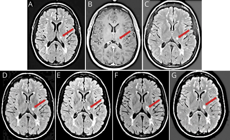
In (A), an axial T2 fluid-attenuated inversion recovery (FLAIR) image
demonstrates a new hyperintense lesion localized to the left thalamus
(red arrows), with periventricular and juxtacortical lesions typical of
MS. In (B), an axial T1 postcontrast scan shows hypointensity of the
left thalamic lesion without contrast enhancement. In (C–G), we
present axial FLAIR images performed serially at 3, 7, 11, 16, and 20
weeks, respectively, after the inception of the right upper extremity
tremor. Over this period, the lesion has slightly increased in size, and
in (F), the lesion takes on a ring configuration with central
hypointensity (red arrows). This lesion failed to exhibit any evidence
of contrast enhancement over the period of surveillance imaging.
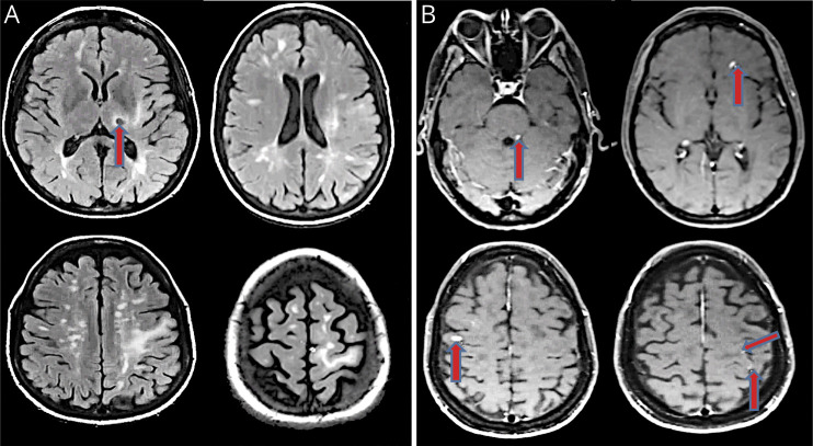
In (A), we present axial fluid-attenuated inversion recovery images revealing
disseminated lesions characteristic for inflammatory demyelination in the
periventribular zones, in the centrum semiovale, in the corona radiata, and
in the cortex and juxtacortical zones, in addition to the previously
identified left thalamic lesion, here exhibiting a ring configuration with
central hypointensity (red arrow). In (B), we present axial T1 postcontrast
images showing various nodular enhancing lesions in left dorsolateral pons
and bilateral frontal lobes and patchy punctate enhancements in left frontal
and parietal white matter (red arrows).
Similar articles
-
Combination of teriflunomide and interferon as follow-up therapy after fingolimod-associated PML.Neurol Neuroimmunol Neuroinflamm. 2020 Dec 3;8(1):e927. doi: 10.1212/NXI.0000000000000927. Print 2021 Jan. Neurol Neuroimmunol Neuroinflamm. 2020. PMID: 33272956 Free PMC article. No abstract available.
-
Treating MS after surviving PML: Discrete strategies for rescue, remission, and recovery patient 2: From the National Multiple Sclerosis Society Case Conference Proceedings.Neurol Neuroimmunol Neuroinflamm. 2020 Dec 15;8(1):e930. doi: 10.1212/NXI.0000000000000930. Print 2021 Jan. Neurol Neuroimmunol Neuroinflamm. 2020. PMID: 33434885 Free PMC article. No abstract available.
-
Fingolimod treatment after natalizumab-related progressive multifocal leukoencephalopathy: three new cases.Mult Scler. 2015 Apr;21(5):671-2. doi: 10.1177/1352458514549823. Epub 2014 Oct 10. Mult Scler. 2015. PMID: 25305251 No abstract available.
-
An updated review of teriflunomide's use in multiple sclerosis.Neurodegener Dis Manag. 2021 Oct;11(5):387-409. doi: 10.2217/nmt-2021-0014. Epub 2021 Sep 16. Neurodegener Dis Manag. 2021. PMID: 34486382 Review.
-
Multiple sclerosis, a treatable disease .Clin Med (Lond). 2017 Dec;17(6):530-536. doi: 10.7861/clinmedicine.17-6-530. Clin Med (Lond). 2017. PMID: 29196354 Free PMC article. Review.
Cited by
-
Recurrent Optic Neuritis and Perineuritis Followed by an Unexpected Discovery: From the National Multiple Sclerosis Society Case Conference Proceedings.Neurol Neuroimmunol Neuroinflamm. 2022 Nov 10;10(1):e200051. doi: 10.1212/NXI.0000000000200051. Print 2023 Jan. Neurol Neuroimmunol Neuroinflamm. 2022. PMID: 36357190 Free PMC article.
-
Multiple Sclerosis Followed by Neuromyelitis Optica Spectrum Disorder: From the National Multiple Sclerosis Society Case Conference Proceedings.Neurol Neuroimmunol Neuroinflamm. 2022 Oct 21;10(1):e200037. doi: 10.1212/NXI.0000000000200037. Print 2023 Jan. Neurol Neuroimmunol Neuroinflamm. 2022. PMID: 36270950 Free PMC article.
-
Escalation to Anti-CD20 Treatment for Multiple Sclerosis Following Natalizumab-Associated Progressive Multifocal Leukoencephalopathy.Neurol Clin Pract. 2024 Oct;14(5):e200330. doi: 10.1212/CPJ.0000000000200330. Epub 2024 Jun 10. Neurol Clin Pract. 2024. PMID: 38919933
References
-
- Yousry TA, Pelletier D, Cadavid D, et al. . Magnetic resonance imaging pattern in natalizumab‐associated progressive multifocal leucoencephalopathy, Ann Neurol 2012;72:779–787. - PubMed
-
- Bloomgren G, Richman S, Hotermans C, et al. . Risk of natalizumab-associated progressive multifocal leukoencephalopathy. N Engl J Med 2012;366:1870–1880. - PubMed
Publication types
MeSH terms
Substances
LinkOut - more resources
Full Text Sources
Medical