. 2021 Feb 22;6(4):e142376.
doi: 10.1172/jci.insight.142376.

A STAT3 inhibitor ameliorates CNS autoimmunity by restoring Teff:Treg balance

Affiliations

A STAT3 inhibitor ameliorates CNS autoimmunity by restoring Teff:Treg balance

Saba I Aqel et al. JCI Insight. .

Abstract

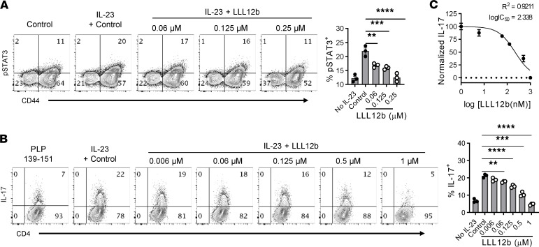
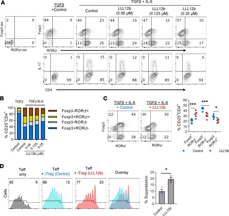
Reestablishing an appropriate balance between T effector cells (Teff) and Tregs is essential for correcting autoimmunity. Multiple sclerosis (MS) is an immune-mediated chronic CNS disease characterized by neuroinflammation, demyelination, and neuronal degeneration, in which the Teff:Treg balance is skewed toward pathogenic Teffs Th1 and Th17 cells. STAT3 is a key regulator of Teff:Treg balance. Using the structure-based design, we have developed a potentially novel small-molecule prodrug LLL12b that specifically inhibits STAT3 and suppresses Th17 differentiation and expansion. Moreover, LLL12b regulates the fate decision between Th17 and Tregs in an inflammatory environment, shifting Th17:Treg balance toward Tregs and favoring the resolution of inflammation. Therapeutic administration of LLL12b after disease onset significantly suppresses disease progression in adoptively transferred, chronic, and relapsing-remitting experimental autoimmune encephalomyelitis. Disease relapses were also significantly suppressed by LLL12b given during the remission phase. Additionally, LLL12b shifts Th17:Treg balance of CD4+ T cells from MS patients toward Tregs and increases Teff sensitivity to Treg-mediated suppression. These data suggest that selective inhibition of STAT3 by the small molecule LLL12b recalibrates the effector and regulatory arms of CD4+ T responses, representing a potentially clinically translatable therapeutic strategy for MS.

Keywords: Autoimmune diseases; Autoimmunity; Demyelinating disorders; Drug therapy; Therapeutics.


Conflict of interest statement

Conflict of interest: The authors have declared that no conflict of interest exists.

Figures

Figure 1
Figure 1. A prodrug of selective STAT3 inhibitor suppresses Th17 development.
(A) Chemical structure of LLL12b. (B) Splenocytes from naive Vα2.3/Vβ8.2 TCR transgenic mice that are specific for MBP Ac1-11 were activated with MBP Ac1-11 plus TGF-β1/IL-6 for 3 days with different concentrations of LLL12b or LLL12. DMSO was used as vehicle control. IL-17 was determined by intracellular staining, gating on CD4+CD44+ T cells. (C and D) Purified CD4+ T cells were activated with αCD3/CD28 plus TGF-β1/IL-6 for 3 days with different concentrations of LLL12b or vehicle control DMSO. IL-17 and RORγt were determined by intracellular staining, gating on CD44+CD4+ cells, and compared with 1-way ANOVA (control, n = 16; 0.125 μM LLL12b, n = 5; 0.25 μM LLL12b, n = 8). (E) Adherent cells from spleens of WT/B6 mice were cultured with different concentrations of LLL12b or vehicle control DMSO for 4–5 hours. The cells were then cultured with purified CD4+ T cells from TCR transgenic 2D2 mice that are specific for MOG 35-55 for 3 days, in the presence of MOG 35-55 and TGF-β/IL-6. IL-17 and RORγt were determined by intracellular staining, gating on CD44+CD4+ cells, and compared with 1-way ANOVA (control, n = 10; 0.125 μM LLL12b, n = 7; 0.5 μM LLL12b, n = 7). Data are represented as mean ± SEM of 3–5 independent experiments. **P < 0.01; ****P < 0.0001.
Figure 2
Figure 2. LLL12b suppresses IL-23–induced expansion of Th17 cells from EAE mice.
(A and B) Splenocytes from immunized SJL/J mice were activated with PLP 139-151 plus IL-23 for 30’ (A) or 3 days (B) with different concentrations of LLL12b. DMSO was used as vehicle control. (A) pSTAT3 was determined by phospho flow cytometry, gating on CD4+ cells, and compared with 1-way ANOVA (n = 3). (B) IL-17 was determined by intracellular staining, gating on CD4+CD44+ T cells. Group means were compared with the control group by 1-way ANOVA (n = 3). (C) Dose response curve of LLL12b concentration and normalized inhibition of % IL-17+CD4+ T cells. Data are represented as mean ± SEM of 3 independent experiments. **P < 0.01; ***P < 0.001; ****P < 0.0001.
Figure 3
Figure 3. LLL12b regulates the fate decision between Th17 and Tregs.
(A and B) Splenocytes from naive Vα2.3/Vβ8.2 TCR transgenic mice were activated with MBP Ac 1-11 plus TGF-β (4 ng/mL) or TGF-β (4 ng/mL)/IL-6 for 3 days with different concentrations of LLL12b. DMSO was used as vehicle control. RORγt, Foxp3, and IL-17 in CD4+ T cells was determined by intracellular staining, gating on CD25+CD4+ T cells (A). (B) Stacked bar graph shows subsets of CD25+CD4+ T cells differentially expressing RORγt and Foxp3 in the groups from the upper panel of A. (C) CD4+ T cells from WT/B6 mice were activated with αCD3/CD28 and TGF-β/IL-6 for 3 days plus LLL12b (0.25 μM) or vehicle control DMSO. RORγt and Foxp3 and in CD4+ T cells was determined by intracellular staining, gating on CD25+CD4+ T cells. Percentage of Foxp3+RORγt, Foxp3+RORγt+, and Foxp3RORγt+ cells in LLL12b or control-treated groups were compared with 1-way ANOVA (n = 5). (D) The cells in C were then mixed with CFSE-labeled splenocytes from naive TCR transgenic 2D2 mice that are specific for MOG 35-55 at a 1:4 ratio and activated with MOG 35-55 for 5 days. CFSE was determined by flow cytometry, gating on CD4+ cells. Percentage of suppression of the proliferation of CFSE-labeled cells were calculated and compared using Mann-Whitney U test (n = 4). Data are represented as mean ± SEM of 3–5 independent experiments. *P < 0.05; ***P < 0.001.
Figure 4
Figure 4. Therapeutic administration of LLL12b significantly suppresses EAE progression.
(AC) Splenocytes from Vα2.3/Vβ8.2 TCR transgenic mice were activated with MBP Ac1-11 plus IL-6 for 3 days (d) and injected into naive B10.PL mice. LLL12b (10 mg/kg) or vehicle control were injected i.p. into mice daily for 7d starting on d10 after adoptive transfer. Peak clinical scores (control, n = 41; LLL12b, n = 34) and AUC (n = 3) were compared (A). On d25 after adoptive transfer, Tregs in the spleens were determined by intracellular staining (control, n = 16; LLL12b, n = 15) (B). Splenocytes (control, n = 10; LLL12b, n = 9) (B) and CNS infiltrating cells (C) were activated with MBP Ac1-11 for 3d (B) or overnight (C). IL-17 was determined by intracellular staining, gating on CD4+CD44+. (D and E) C57BL/6 mice were immunized with MOG 35-55 and treated with LLL12b (or control) as described in A starting on d14 after immunization. On d29 after immunization, splenocytes were activated with MOG 35-55 for 3d. IL-17 in supernatant was determined by ELISA (control, n = 16; LLL12b, n = 17) (E). (FH) SJL/J mice were immunized with PLP 139-151 and treated with LLL12b (or control) as described in A starting on d9 (F) or d22 (G and H). Peak clinical scores (n = 34) and normalized AUC (n = 3) were compared (G). On d33 after immunization, splenocytes of mice in G were activated with PLP 139-151 for 3d. IL-17 in supernatant was determined by ELISA (LLL12b, n = 13; control, n = 15). EAE clinical scores were compared with Mann-Whitney U test. Bar graphs were compared with unpaired Student’s t test. Data represent mean ± SEM of 3–5 independent experiments. *P < 0.05; **P < 0.01; ***P < 0.001; ****P < 0.0001.
Figure 5
Figure 5. LLL12b restores Th17.
Treg balance of CD4+ T cells from MS patients. (AL) PBMCs from treatment-naive MS patients (n = 22) were activated with αCD3/CD28 plus TGF-β/IL-6 for 3 days with LLL12b. DMSO was used as vehicle control. (AD) IL-17 in supernatant of 1 patient sample was determined by ELISA and compared with 1-way ANOVA (A). IL-17 in LLL12b (0.125 μM) group was compared with control group of the same patient using Wilcoxon matched-pairs signed rank test (B). The percentage of decrease of IL-17 was calculated (C), and the patients in different ranges were shown in a pie chart (D). (EH) Tregs (FoxP3+CD25+) were determined by intracellular staining, gating on CD45RA+CD4+ cells (E), and compared between LLL12b- (0.125 μM) and control-treated groups of the same patient using Wilcoxon matched-pairs signed rank test (F). The percentage of increase of Tregs was calculated (G), and the patients in different ranges were shown in a pie chart (H). (IK) IL-17/Treg ratio of each patient was calculated and compared between LLL12b and control groups with Wilcoxon matched-pairs signed rank test (I). The percentage of decrease of IL-17/Treg ratio was calculated (J) and shown in a pie chart (K). (L) A nonparametric Pearson correlation test was used to analyze the degree of relatedness between percent increase of Treg and percent decrease of IL-17. (M) PBMCs from MS patients (n = 3) were activated with αCD3/CD28 under iTreg differentiation condition for 3 days. CFSE-labeled PBMCs from the same 3 patients were cultured with LLL12b (0.25 μM) or vehicle control DMSO for 2 hours, washed and mixed with iTregs cultured cells (Teff:Treg = 16:1), followed by activation with αCD3/CD28 for 5 days. CFSE in CD4+ T cells was determined by flow cytometry. The percentage of suppression was calculated and compared with a paired Student’s t test. **P < 0.01; ****P < 0.0001.

References

    1. Wallin MT, et al. The prevalence of MS in the United States: a population-based estimate using health claims data. Neurology. 2019;92(10):1029–1040. - PMC - PubMed
    1. Arellano B, et al. Regulatory T cell-based therapies for autoimmunity. Discov Med. 2016;22(119):73–80. - PMC - PubMed
    1. Yang Y, et al. T-bet is essential for encephalitogenicity of both Th1 and Th17 cells. J Exp Med. 2009;206(7):1549–1564. doi: 10.1084/jem.20082584. - DOI - PMC - PubMed
    1. Das J, et al. Transforming growth factor beta is dispensable for the molecular orchestration of Th17 cell differentiation. J Exp Med. 2009;206(11):2407–2416. doi: 10.1084/jem.20082286. - DOI - PMC - PubMed
    1. Ghoreschi K, et al. Generation of pathogenic T(H)17 cells in the absence of TGF-β signalling. Nature. 2010;467(7318):967–971. doi: 10.1038/nature09447. - DOI - PMC - PubMed

Publication types

MeSH terms