. 2021 Jan 7;16(1):e0243788.
doi: 10.1371/journal.pone.0243788. eCollection 2021.

CX3CR1 is a prerequisite for the development of cardiac hypertrophy and left ventricular dysfunction in mice upon transverse aortic constriction

Affiliations

CX3CR1 is a prerequisite for the development of cardiac hypertrophy and left ventricular dysfunction in mice upon transverse aortic constriction

Christina Katharina Weisheit et al. PLoS One. .

Abstract

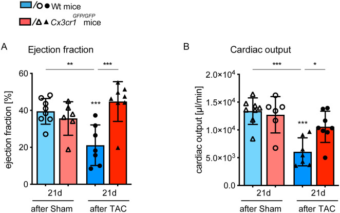
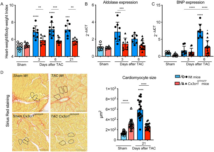
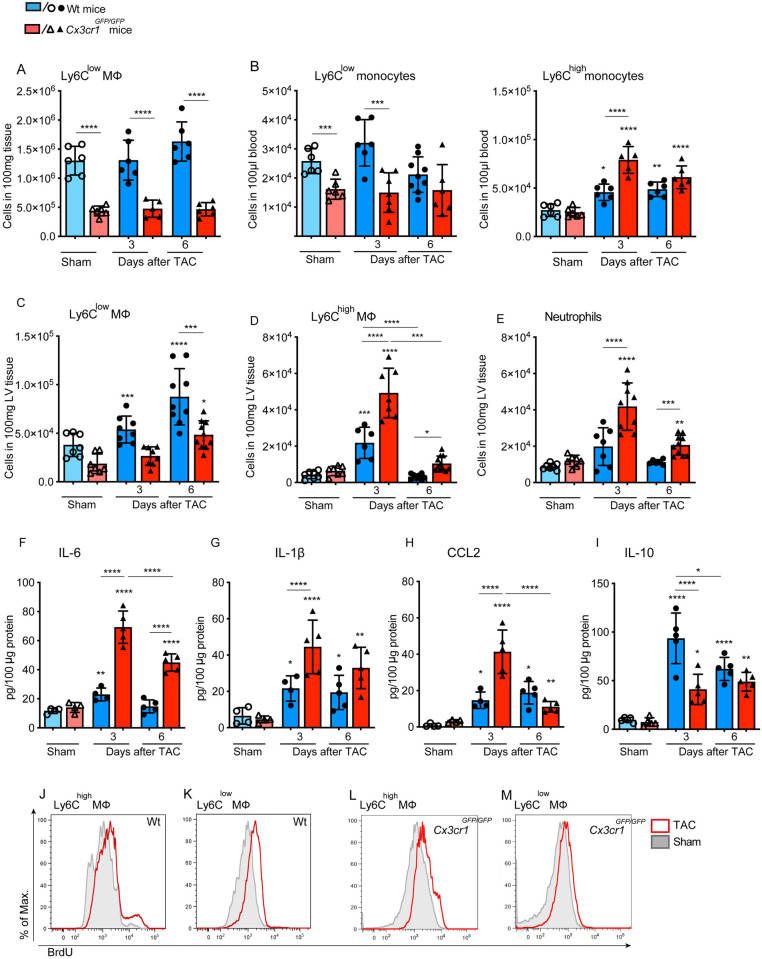
The CX3CL1/CX3CR1 axis mediates recruitment and extravasation of CX3CR1-expressing subsets of leukocytes and plays a pivotal role in the inflammation-driven pathology of cardiovascular disease. The cardiac immune response differs depending on the underlying causes. This suggests that for the development of successful immunomodulatory therapy in heart failure due to chronic pressure overload induced left ventricular (LV) hypertrophy, the underlying immune patterns must be examined. Here, the authors demonstrate that Fraktalkine-receptor CX3CR1 is a prerequisite for the development of cardiac hypertrophy and left ventricular dysfunction in a mouse model of transverse aortic constriction (TAC). The comparison of C57BL/6 mice with CX3CR1 deficient mice displayed reduced LV hypertrophy and preserved cardiac function in response to pressure overload in mice lacking CX3CR1. Moreover, the normal immune response following TAC induced pressure overload which is dominated by Ly6Clow macrophages changed to an early pro-inflammatory immune response driven by neutrophils, Ly6Chigh macrophages and altered cytokine expression pattern in CX3CR1 deficient mice. In this early inflammatory phase of LV hypertrophy Ly6Chigh monocytes infiltrated the heart in response to a C-C chemokine ligand 2 burst. CX3CR1 expression impacts the immune response in the development of LV hypertrophy and its absence has clear cardioprotective effects. Hence, suppression of CX3CR1 may be an important immunomodulatory therapeutic target to ameliorate pressure-overload induced heart failure.


Conflict of interest statement

This study was supported by the Flow-cytometry Core Facility and by the House for Experimental Therapy (HET) of the medical faculty of Bonn University. C. K. W. and L. E. were supported by the Else Kröner-Forschungskolleg Bonn and BONFOR in Bonn; C. K. W., S. Z. and C. K. are funded by the Deutsche Forschungsgemeinschaft (DFG, German Research Foundation) – Grant No. 397484323 – Project number 426093965. C. K. and S.Z. are members of the excellence cluster ‘‘ImmunoSensation’’ at Bonn University. There are no patents, products in development or marketed products to declare. This does not alter our adherence to PLOS ONE policies on sharing data and materials.

Figures

Fig 1
Fig 1. Cx3cr1GFP/GFP mice exhibit a cardioprotective phenotype in response to chronic pressure overload.
Using pressure volume measurement, the cardiac function of Cx3cr1GFP/GFP mice and Wt mice was evaluated 21 days after TAC or sham operation. (A, B) Ejection fraction and cardiac output are depicted as plots, more information is provided in Table 1. N = 6–8 mice/group; * above individual columns indicate significant differences between TAC and respective sham group; *P <0.05, **P<0.01, *** P<0.001.
Fig 2
Fig 2. CX3CR1 expression modulates the development of LV hypertrophy in response to pressure overload.
(A) Heart-Weight/Body-Weight-Index of Wt and Cx3cr1GFP/GFP mice calculated 3, 6 and 21 days after transverse aortic constriction (TAC) and sham operation; n = 7–9 mice/group. (B) Aldolase mRNA expression pattern in LV tissue determined 3 and 6 days after TAC and sham operation in Wt and Cx3cr1GFP/GFP mice; n = 6–14 mice/group. (C) BNP mRNA expression pattern in LV tissue determined 3 and 6 days after TAC and sham operation in Wt and Cx3cr1GFP/GFP mice; n = 6–14 mice/group. (D) Representative sections of the left ventricle stained with sirious red of Wt and Cx3cr1GFP/GFP mice 21 days after TAC or Sham operation to determine cardiomyocyte size. Dashed lines highlight exemplary individual cardiomyocytes; scale bar indicating 50μm. Quantification of cardiomyocyte size by area planimetry; n = 5–6 mice/group, 4 slides per mouse were randomly analyzed by manual cell count of at least 50 cells/slide and mean was used for statistical analysis. * above individual columns indicate significant differences between TAC and respective sham group; **P<0.01, *** P<0.001, ****p < 0.0001.
Fig 3
Fig 3. CX3CR1 deficiency drives the immune response to a pro-inflammatory phenotype during chronic pressure overload.
Quantification of Ly6Clow F4/80+ macrophages in the spleen and of (B) Ly6Clow and Ly6Chigh CD115+ monocytes in the blood of Wt and Cx3cr1GFP/GFP mice determined by flow cytometry 3 and 6 days after TAC and sham ioperation. (C-E) Number of neutrophils, Ly6Clow and Ly6Chigh macrophages in the LV tissue was determined by flow cytometry analysis in Cx3cr1GFP/GFP and Wt mice 3 and 6 days after TAC and sham operation. The particular cell subsets were defined as follows: neutrophils as CD45+ F4/80- Ly6G+, macrophages as CD45+ F4/80+ Ly6G-; Ly6Chigh and Ly6Clow macrophages were further discriminated according to the respective Ly6C surface expression; (F-I) Concentrations of the pro-inflammatory cytokines IL6 and IL-1β, the chemokine MCP1 and IL-10 were determined in LV tissue homogenates at day 3 and 6 after surgical intervention in Cx3cr1GFP/GFP and Wt mice using ELISA assays. (J-M) BrdU content of cardiac Ly6Chigh and Ly6Clow macrophages was determined by flow cytometry analysis at day 3 after TAC. BrdU was administered 18h before analysis. Histograms are based on concetenated plots of 4 mice. N = 6–8 mice/group; * above individual columns indicate significant differences between TAC and respective sham group; *P <0.05, **P<0.01, *** P<0.001, ****p < 0.0001.

References

    1. Koga K, Kenessey A, Ojamaa K. Macrophage migration inhibitory factor antagonizes pressure overload-induced cardiac hypertrophy. American journal of physiology Heart and circulatory physiology. 2013;304(2):H282–93. Epub 2012/11/13. 10.1152/ajpheart.00595.2012 . - DOI - PubMed
    1. Duerr GD, Heinemann JC, Kley J, Eichhorn L, Frede S, Weisheit C, et al. Myocardial maladaptation to pressure overload in CB2 receptor-deficient mice. J Mol Cell Cardiol. 2019;133:86–98. Epub 2019/06/11. 10.1016/j.yjmcc.2019.06.003 . - DOI - PubMed
    1. Velten M, Duerr GD, Pessies T, Schild J, Lohner R, Mersmann J, et al. Priming with synthetic oligonucleotides attenuates pressure overload-induced inflammation and cardiac hypertrophy in mice. Cardiovasc Res. 2012;96(3):422–32. Epub 2012/09/15. . - PubMed
    1. Massie BM. Novel targets for the treatment of heart failure: perspectives from a heart failure clinician and trialist. J Mol Cell Cardiol. 2011;51(4):438–40. Epub 2011/05/10. 10.1016/j.yjmcc.2011.03.016 . - DOI - PubMed
    1. Kurrelmeyer K, Kalra D, Bozkurt B, Wang F, Dibbs Z, Seta Y, et al. Cardiac remodeling as a consequence and cause of progressive heart failure. Clinical cardiology. 1998;21(12 Suppl 1):I14–9. Epub 1998/12/16. 10.1002/clc.4960211304 . - DOI - PMC - PubMed

Publication types

MeSH terms