Review
. 2021 Mar;24 Suppl 1(Suppl 1):16-33.
doi: 10.1111/vop.12858. Epub 2021 Jan 7.

Looking into the future: Gene and cell therapies for glaucoma

Affiliations
Review

Looking into the future: Gene and cell therapies for glaucoma

András M Komáromy et al. Vet Ophthalmol. 2021 Mar.

Abstract

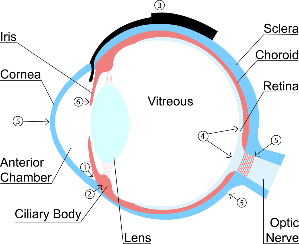
Glaucoma is a complex group of optic neuropathies that affects both humans and animals. Intraocular pressure (IOP) elevation is a major risk factor that results in the loss of retinal ganglion cells (RGCs) and their axons. Currently, lowering IOP by medical and surgical methods is the only approved treatment for primary glaucoma, but there is no cure, and vision loss often progresses despite therapy. Recent technologic advances provide us with a better understanding of disease mechanisms and risk factors; this will permit earlier diagnosis of glaucoma and initiation of therapy sooner and more effectively. Gene and cell therapies are well suited to target these mechanisms specifically with the potential to achieve a lasting therapeutic effect. Much progress has been made in laboratory settings to develop these novel therapies for the eye. Gene and cell therapies have already been translated into clinical application for some inherited retinal dystrophies and age-related macular degeneration (AMD). Except for the intravitreal application of ciliary neurotrophic factor (CNTF) by encapsulated cell technology for RGC neuroprotection, there has been no other clinical translation of gene and cell therapies for glaucoma so far. Possible application of gene and cell therapies consists of long-term IOP control via increased aqueous humor drainage, including inhibition of fibrosis following filtration surgery, RGC neuroprotection and neuroregeneration, modification of ocular biomechanics for improved IOP tolerance, and inhibition of inflammation and neovascularization to prevent the development of some forms of secondary glaucoma.

Keywords: cell therapy; gene therapy; glaucoma; intraocular pressure (IOP); neuroprotection; ocular biomechanics.


Figures

Figure 1.
Figure 1.
Schematic cross-section of an eye showing potential targets for gene and cell glaucoma therapies: (1) Increased aqueous humor outflow, (2) decreased aqueous humor production, (3) preventing gonioimplant bleb fibrosis, (4) RGC neuroprotection and neuroregeneration, (5) modification of biomechanical properties of sclera, lamina cribrosa, and cornea, and (6) inhibition of inflammation and PIFVM formation.
Figure 2.
Figure 2.
Schematic cross-section of an eye showing possible methods for intraocular administration of gene and cell therapies for glaucoma. (1) Gene therapy vectors or cells for the treatment of the aqueous humor outflow pathways within the ICA are administered by intracameral injection. (2) Injections through the pars plana of the ciliary body into the anterior vitreous are used to target the ciliary body epithelium; reagents will also reach the anterior chamber and aqueous humor outflow pathways. (3) Gene therapy vectors or cells for neuroprotective therapy are injected through the pars plana into the posterior vitreous, close to the surface of the retina and ONH. (4) Capsules with genetically modified cells, that release neuroprotective reagents, are attached to the sclera at the pars plana and reach into the anterior vitreous. (5) Transplantation of RGCs to restore vision are injected into the posterior vitreous or (not shown) underneath the retina’s inner limiting membrane. If successful, the cells embed in the retina and form axons that extend through the optic nerve and connect with targets in the brain.
Figure 3.
Figure 3.
Targeting of GFP transgene expression to the canine anterior ocular segment by intravitreal injection of capsid mutated AAV2 (triple Y-F+T-V). (A) Fluorescent gonioscopic imaging showing fluorescence within the ciliary cleft 6 weeks post intravitreal AAV injection. Note the pectinate ligament fibrils demonstrated by the dark bands crossing the region of fluorescence. (B) Immunohistochemical labeling of cryosections of the canine anterior segment shows GFP within the trabecular meshwork (arrow). (C) GFP expression is also seen within the ciliary body epithelium (arrow). GFP, green fluorescent protein; triple Y-F + T-V, AAV2 based capsid was mutated by substitution of three surface-exposed capsid tyrosine (Y) residues with phenylalanine (F) and one threonine (T) residue with valine (V). Scale bar, 100 μm. Boyd et al. 2016, with permission.
Figure 4.
Figure 4.
Transduction of canine RGC following intravitreal injection of capsid mutated AAV2 (triple Y-F + T-V) with GFP transgene. (A) Representative confocal scanning laser ophthalmoscopy image obtained at 5 weeks post injection demonstrated widespread GFP fluorescence. (B) Immunohistochemical labeling of retinal cryosection with neuronal nuclei (NeuN) antibody (red) to label RGCs demonstrates a high number of cells colabeling with GFP (green). Cell nuclei are shown in blue with DAPI. DAPI, 4’,6-diamidino-2-phenylindole; GCL, ganglion cell layer; GFP, green fluorescent protein; INL, inner nuclear layer; ONL, outer nuclear layer; triple Y-F+T-V, AAV2 based capsid was mutated by substitution of three surface-exposed capsid tyrosine (Y) residues with phenylalanine (F) and one threonine (T) residue with valine (V). Scale bar, 50 μm. Boyd et al. 2016, with permission.

Similar articles

Cited by

References

    1. Weinreb RN, Aung T, Medeiros FA. The pathophysiology and treatment of glaucoma: a review. JAMA. 2014;311(18):1901–1911. - PMC - PubMed
    1. Komaromy AM, Bras D, Esson DW, et al. The future of canine glaucoma therapy. Veterinary ophthalmology. 2019;22(5):726–740. - PMC - PubMed
    1. Gelatt KN, MacKay EO. Prevalence of the breed-related glaucomas in pure-bred dogs in North America. Veterinary ophthalmology. 2004;7(2):97–111. - PubMed
    1. Gelatt KN, MacKay EO. Secondary glaucomas in the dog in North America. Veterinary ophthalmology. 2004;7(4):245–259. - PubMed
    1. Strom AR, Hassig M, Iburg TM, Spiess BM. Epidemiology of canine glaucoma presented to University of Zurich from 1995 to 2009. Part 2: secondary glaucoma (217 cases). Veterinary ophthalmology. 2011;14(2):127–132. - PubMed