

Can SARS-CoV-2 infect the central nervous system via the olfactory bulb or the blood-brain barrier?
- PMID: 33412255
- PMCID: PMC7836942
- DOI: 10.1016/j.bbi.2020.12.031
Can SARS-CoV-2 infect the central nervous system via the olfactory bulb or the blood-brain barrier?
Abstract
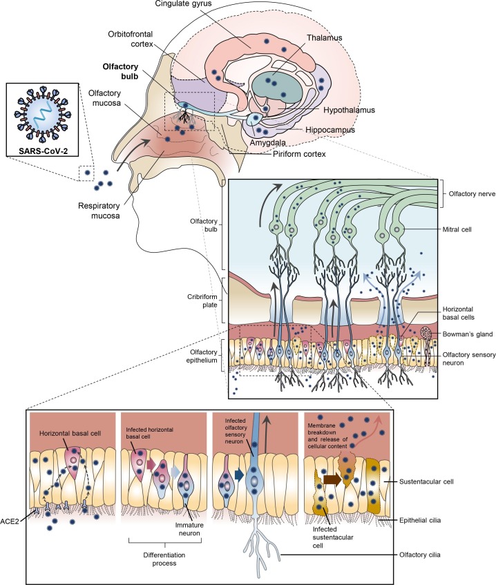
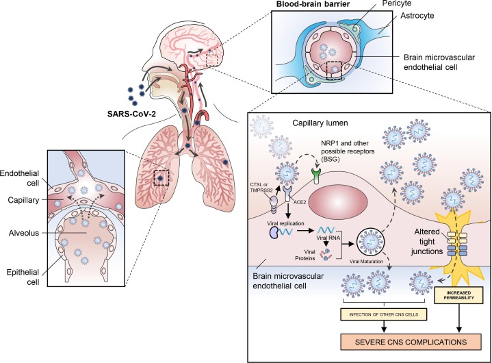
Severe acute respiratory syndrome coronavirus 2 (SARS-CoV-2) emerged in Wuhan, China in December 2019. On February 11, the World Health Organization (WHO) announced the name for the new illness caused by SARS-CoV-2: COVID-19. By March 11, the outbreak of COVID-19 was declared a pandemic by the WHO. This virus has extensively altered daily life for many across the globe, while claiming hundreds of thousands of lives. While fundamentally a respiratory illness, many infected individuals experience symptoms that involve the central nervous system (CNS). It is likely that many of these symptoms are the result of the virus residing outside of the CNS. However, the current evidence does indicate that the SARS-CoV-2 virus can use olfactory neurons (or other nerve tracts) to travel from the periphery into the CNS, and that the virus may also enter the brain through the blood-brain barrier (BBB). We discuss how the virus may use established infection mechanisms (ACE2, NRP1, TMPRSS2, furin and Cathepsin L), as well mechanisms still under consideration (BASIGIN) to infect and spread throughout the CNS. Confirming the impact of the virus on the CNS will be crucial in dealing with the long-term consequences of the epidemic.
Published by Elsevier Inc.
Figures
References
-
- Arslan O. second ed. CRC Press; 2014. Olfactory System. Neuroanatomical Basis of Clinical Neurology; pp. 377–386.
-
- Atlas, H. P., 2020. “The Human Protein Atlas.” Retrieved Sept 3, 2020, from https://www.proteinatlas.org/.
-
- Baig A.M., Khaleeq A., Ali U., Syeda H. Evidence of the COVID-19 Virus Targeting the CNS: tissue distribution, host-virus interaction, and proposed neurotropic mechanisms. ACS Chem. Neurosci. 2020;11(7):995–998. - PubMed
-
- Barnes B.J., Adrover J.M., Baxter-Stoltzfus A., Borczuk A., Cools-Lartigue J., Crawford J.M., Dassler-Plenker J., Guerci P., Huynh C., Knight J.S., Loda M., Looney M.R., McAllister F., Rayes R., Renaud S., Rousseau S., Salvatore S., Schwartz R.E., Spicer J.D., Yost C.C., Weber A., Zuo Y., Egeblad M. Targeting potential drivers of COVID-19: neutrophil extracellular traps. J. Exp. Med. 2020;217(6) - PMC - PubMed
Publication types
MeSH terms
LinkOut - more resources
Full Text Sources
Other Literature Sources
Medical
Miscellaneous
