

Low-Intensity Pulsed Ultrasound Promotes Autophagy-Mediated Migration of Mesenchymal Stem Cells and Cartilage Repair
- PMID: 33412895
- PMCID: PMC7797574
- DOI: 10.1177/0963689720986142
Low-Intensity Pulsed Ultrasound Promotes Autophagy-Mediated Migration of Mesenchymal Stem Cells and Cartilage Repair
Abstract
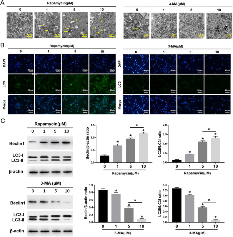
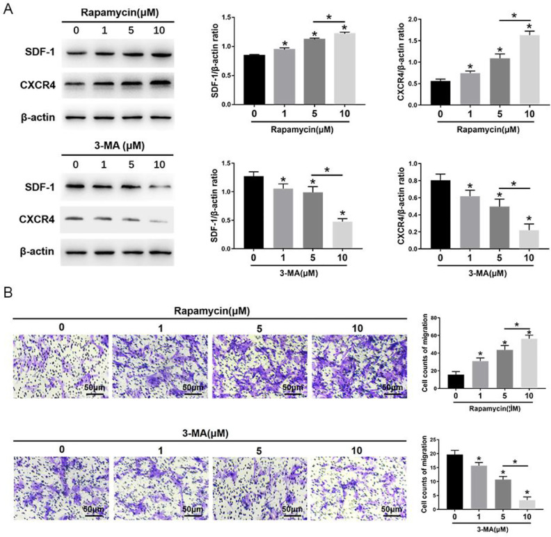
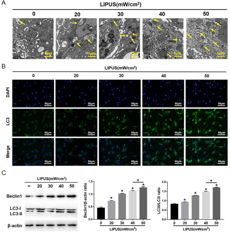
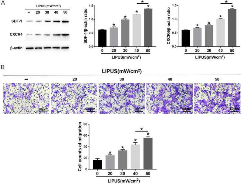
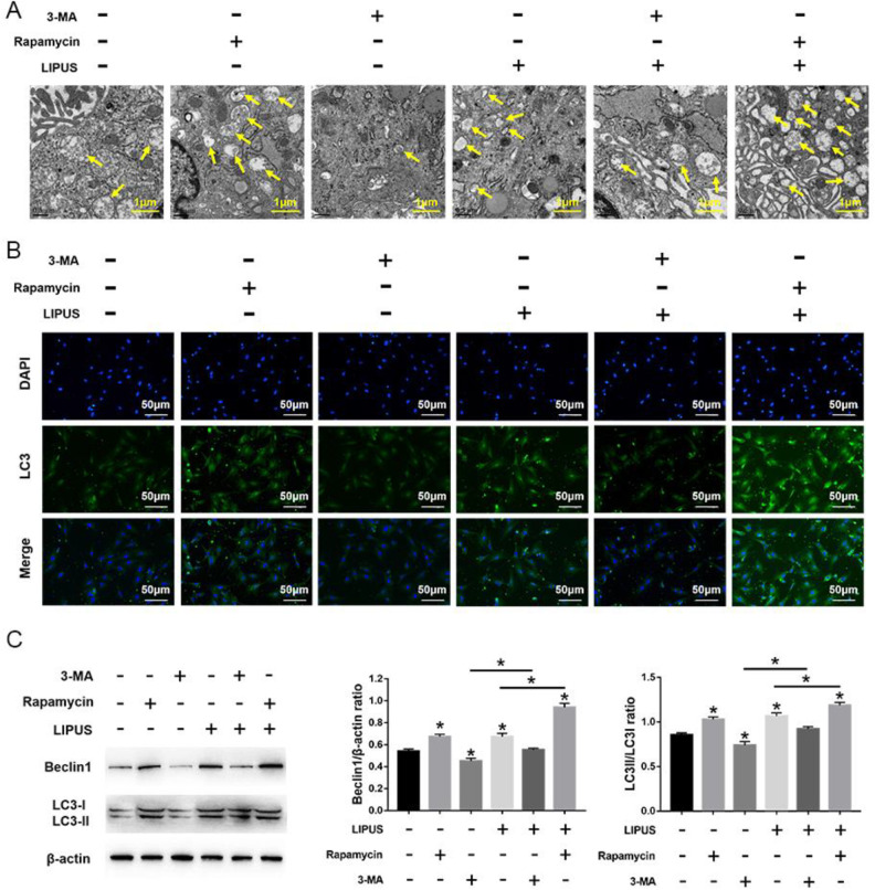
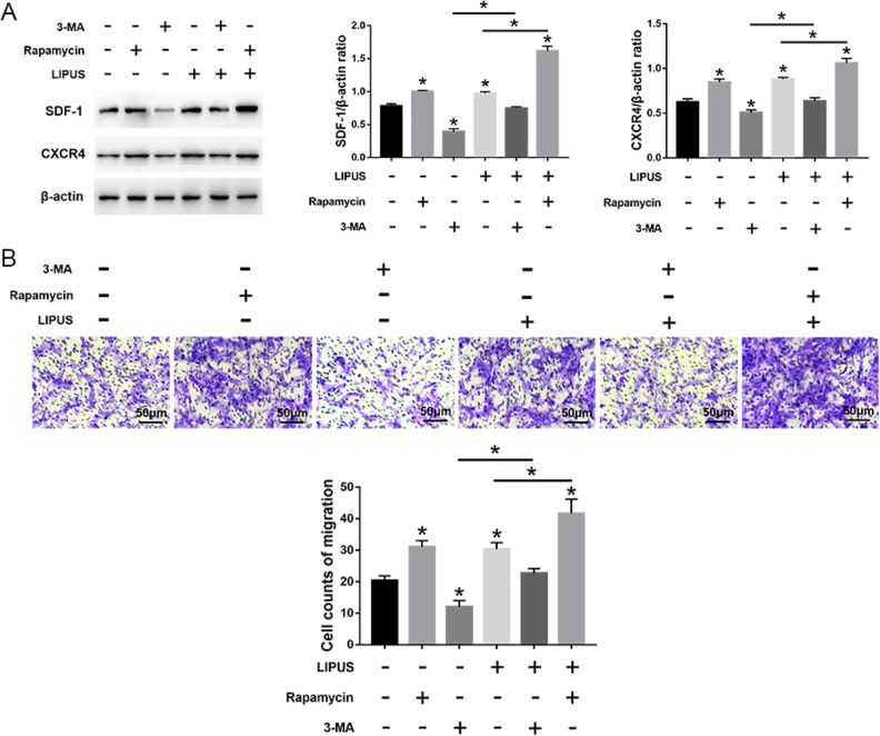
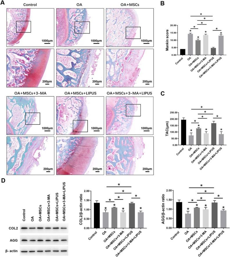
Mesenchymal stem cell (MSC) migration is promoted by low-intensity pulsed ultrasound (LIPUS), but its mechanism is unclear. Since autophagy is known to regulate cell migration, our study aimed to investigate if LIPUS promotes the migration of MSCs via autophagy regulation. We also aimed to investigate the effects of intra-articular injection of MSCs following LIPUS stimulation on osteoarthritis (OA) cartilage. For the in vitro study, rat bone marrow-derived MSCs were treated with an autophagy inhibitor or agonist, and then they were stimulated by LIPUS. Migration of MSCs was detected by transwell migration assays, and stromal cell-derived factor-1 (SDF-1) and C-X-C chemokine receptor type 4 (CXCR4) protein levels were quantified. For the in vivo study, a rat knee OA model was generated and treated with LIPUS after an intra-articular injection of MSCs with autophagy inhibitor added. The cartilage repair was assessed by histopathological analysis and extracellular matrix protein expression. The in vitro results suggest that LIPUS increased the expression of SDF-1 and CXCR4, and it promoted MSC migration. These effects were inhibited and enhanced by autophagy inhibitor and agonist, respectively. The in vivo results demonstrate that LIPUS significantly enhanced the cartilage repair effects of MSCs on OA, but these effects were blocked by autophagy inhibitor. Our results suggest that the migration of MSCs was enhanced by LIPUS through the activation autophagy, and LIPUS improved the protective effect of MSCs on OA cartilage via autophagy regulation.
Keywords: autophagy; mesenchymal stem cell; migration; ultrasound.
Conflict of interest statement
Figures
Similar articles
-
Advances in the application of low-intensity pulsed ultrasound to mesenchymal stem cells.Stem Cell Res Ther. 2022 May 26;13(1):214. doi: 10.1186/s13287-022-02887-z. Stem Cell Res Ther. 2022. PMID: 35619156 Free PMC article. Review.
-
Low-Intensity Pulsed Ultrasound Enhances the Efficacy of Bone Marrow-Derived MSCs in Osteoarthritis Cartilage Repair by Regulating Autophagy-Mediated Exosome Release.Cartilage. 2022 Apr-Jun;13(2):19476035221093060. doi: 10.1177/19476035221093060. Cartilage. 2022. PMID: 35438034 Free PMC article.
-
Effects of Low-Intensity Pulsed Ultrasound on the Migration and Homing of Human Amnion-Derived Mesenchymal Stem Cells to Ovaries in Rats With Premature Ovarian Insufficiency.Cell Transplant. 2022 Jan-Dec;31:9636897221129171. doi: 10.1177/09636897221129171. Cell Transplant. 2022. PMID: 36282038 Free PMC article.
-
Low-intensity pulsed ultrasound promotes mesenchymal stem cell transplantation-based articular cartilage regeneration via inhibiting the TNF signaling pathway.Stem Cell Res Ther. 2023 Apr 17;14(1):93. doi: 10.1186/s13287-023-03296-6. Stem Cell Res Ther. 2023. PMID: 37069673 Free PMC article.
-
The Role of Low-Intensity Pulsed Ultrasound on Cartilage Healing in Knee Osteoarthritis: A Review.PM R. 2017 Dec;9(12):1268-1277. doi: 10.1016/j.pmrj.2017.05.008. Epub 2017 Jun 9. PM R. 2017. PMID: 28606838 Review.
Cited by
-
A Novel Autophagy-Related Marker for Improved Differential Diagnosis of Rheumatoid Arthritis and Osteoarthritis.Front Genet. 2021 Oct 12;12:743560. doi: 10.3389/fgene.2021.743560. eCollection 2021. Front Genet. 2021. PMID: 34712268 Free PMC article.
-
Advances in the application of low-intensity pulsed ultrasound to mesenchymal stem cells.Stem Cell Res Ther. 2022 May 26;13(1):214. doi: 10.1186/s13287-022-02887-z. Stem Cell Res Ther. 2022. PMID: 35619156 Free PMC article. Review.
-
Low-Intensity Pulsed Ultrasound: A Physical Stimulus with Immunomodulatory and Anti-inflammatory Potential.Ann Biomed Eng. 2024 Aug;52(8):1955-1981. doi: 10.1007/s10439-024-03523-y. Epub 2024 Apr 29. Ann Biomed Eng. 2024. PMID: 38683473 Review.
-
Key mediators of the efficacy of mesenchymal stem cells on in vivo disease models.Cell Transplant. 2025 Jan-Dec;34:9636897251348566. doi: 10.1177/09636897251348566. Epub 2025 Jun 25. Cell Transplant. 2025. PMID: 40560652 Free PMC article. Review.
-
Exploration of the optimal retention method in vivo for stem cell therapy: Low-intensity ultrasound preconditioning.Regen Ther. 2025 Apr 30;29:484-492. doi: 10.1016/j.reth.2025.04.012. eCollection 2025 Jun. Regen Ther. 2025. PMID: 40390863 Free PMC article.
References
-
- Wang Y, Yuan M, Guo Q, Lu S, Peng J. Mesenchymal stem cells for treating articular cartilage defects and osteoarthritis. Cell Transplant. 2015;24(9):1661–1678. - PubMed
-
- Short B, Brouard N, Occhiodoro-Scott T, Ramakrishnan A, Simmons PJ. Mesenchymal stem cells. Arch Med Res. 2003;34(6):565–571. - PubMed
-
- Pittenger MF, Mackay AM, Beck SC, Jaiswal RK, Douglas R, Mosca JD, Moorman MA, Simonetti DW, Craig S, Marshak DR. Multilineage potential of adult human mesenchymal stem cells. Science. 1999; 284(5411):143–147. - PubMed
-
- Quintavalla J, Uziel-Fusi S, Yin J, Boehnlein E, Pastor G, Blancuzzi V, Singh HN, Kraus KH, O’Byrne E, Pellas TC. Fluorescently labeled mesenchymal stem cells (mscs) maintain multilineage potential and can be detected following implantation into articular cartilage defects. Biomaterials. 2002;23(1):109–119. - PubMed
-
- Pittenger MF, Martin BJ. Mesenchymal stem cells and their potential as cardiac therapeutics. Circ Res. 2004;95(1):9–20. - PubMed
Publication types
MeSH terms
LinkOut - more resources
Full Text Sources
Other Literature Sources
Medical
