Case Reports
. 2021 Jun 15;38(12):1620-1631.
doi: 10.1089/neu.2020.7373. Epub 2021 Feb 18.

Tractography-Pathology Correlations in Traumatic Brain Injury: A TRACK-TBI Study

Collaborators, Affiliations
Case Reports

Tractography-Pathology Correlations in Traumatic Brain Injury: A TRACK-TBI Study

Amber L Nolan et al. J Neurotrauma. .

Abstract

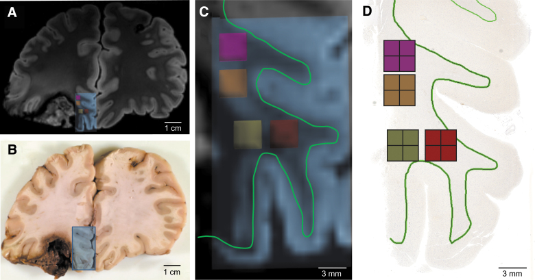
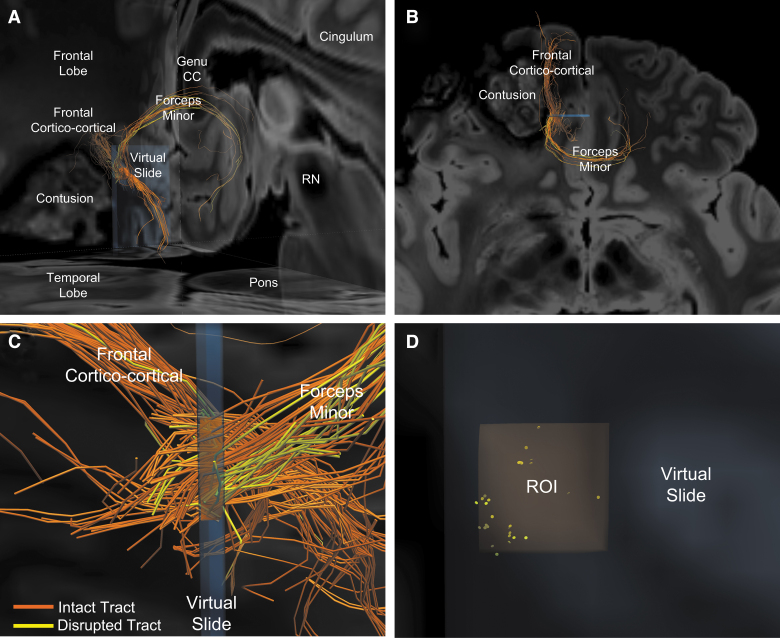
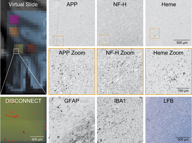
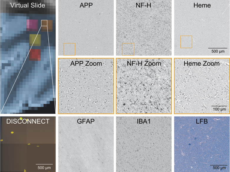
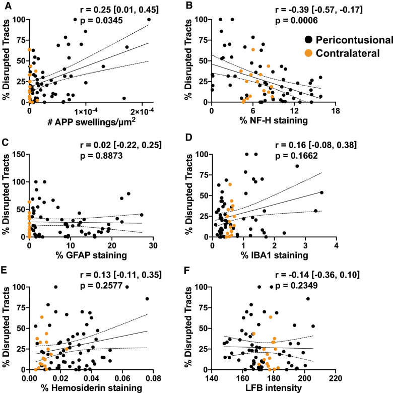
Diffusion tractography magnetic resonance imaging (MRI) can infer changes in network connectivity in patients with traumatic brain injury (TBI), but the pathological substrates of disconnected tracts have not been well defined because of a lack of high-resolution imaging with histopathological validation. We developed an ex vivo MRI protocol to analyze tract terminations at 750-μm isotropic resolution, followed by histopathological evaluation of white matter pathology, and applied these methods to a 60-year-old man who died 26 days after TBI. Analysis of 74 cerebral hemispheric white matter regions revealed a heterogeneous distribution of tract disruptions. Associated histopathology identified variable white matter injury with patchy deposition of amyloid precursor protein (APP), loss of neurofilament-positive axonal processes, myelin dissolution, astrogliosis, microgliosis, and perivascular hemosiderin-laden macrophages. Multiple linear regression revealed that tract disruption strongly correlated with the density of APP-positive axonal swellings and neurofilament loss. Ex vivo diffusion MRI can detect tract disruptions in the human brain that reflect axonal injury.

Keywords: MRI; contusion; neuropathology; tractography; traumatic axonal injury; traumatic brain injury.


Conflict of interest statement

Dr. Fischl has a financial interest in CorticoMetrics, a company whose medical pursuits focus on brain imaging and measurement technologies. His interests were reviewed and are managed by Massachusetts General Hospital and Partners HealthCare in accordance with their conflict of interest policies.

The United States Department of Energy supports Dr. Manley for a precision medicine collaboration. One Mind has provided funding for TRACK-TBI patient stipends and support to clinical sites. Dr. Manley has received an unrestricted gift from the NFL to the UCSF Foundation to support research efforts of the TRACK-TBI NETWORK. Dr. Manley has also received funding from NeuroTrauma Sciences LLC to support TRACK-TBI data curation efforts. Additionally, Abbott Laboratories has provided funding for add-in TRACK-TBI clinical studies.

Ms. Markowitz receives salary support from the United States Department of Energy precision medicine collaboration and One Mind.

Figures

FIG. 1.
FIG. 1.
Coregistration of pericontusional regions for analysis in ex vivo imaging and histologic sections. (A) Coronal section of MRI at the level of the left inferior frontal contusion. Blue box indicates area sampled for histology. (B) Gross pathology of the same region. Blue box indicates the tissue block taken for histology. (C) Magnification of area taken for histologic sampling from (A). Small pink, orange, yellow and red boxes indicate regions of analysis for tractography, which were co-registered to the histology by mapping their spatial relationship to the gray-white junction (green line). (D) Image of tissue section with pink, orange, yellow and red boxes indicating regions of analysis for histology. A green outline of the gray-white junction is shown in (D) to match the green outline shown in (C). MRI, magnetic resonance imaging.
FIG. 2.
FIG. 2.
Representative image of intact and disrupted tracts passing through a pericontusional region of interest (ROI). (A) Postero-lateral oblique perspective of tracts passing through the orange ROI from Figure 1. Two bundles of tracts are identified: the forceps minor crossing the genu of the corpus callosum and a frontal cortico-cortical white matter bundle. (B) Higher magnification of image in (A), shown from a superior perspective. Orange indicates intact tracts, whereas yellow indicates disconnected tracts that terminate in the orange ROI. (C) Left lateral perspective of the orange ROI within the blue virtual slide. Intact (orange) and disconnected (yellow) tracts are seen within forceps minor and the frontal cortico-cortical bundles, with the former showing a higher percentage of disrupted tracts. (D) Anterior view of disconnected tract endpoints (yellow discs) within the orange ROI reveals that the tracts tend to be disconnected in small clusters. CC, corpus callosum; RN, red nucleus.
FIG. 3.
FIG. 3.
Representative histology of pericontusional region of interest with numerous disrupted tracts. The virtual slide demonstrates a pericontusional region on MRI and the associated disrupted tracts by DISCONNECT analysis; disrupted tracts are shown in orange. In the same region of interest, images are shown for immunohistochemistry of amyloid precursor protein (APP), neurofilament (NF-H), glial fibrillary acidic protein (GFAP), and ionized calcium-binding adaptor molecule 1 (IBA1). Hemosiderin (Heme) deposition is identified on a counterstained slide. A Luxol fast blue (LFB) stain indicative of myelin is also displayed. Substantial deposition of APP and a lack of fine background processes with only course sparse swollen segments with NF-H is seen, consistent with the large number of disrupted tracts identified in this region. MRI, magnetic resonance imaging.
FIG. 4.
FIG. 4.
Representative histology of contralateral region of interest with few disrupted tracts. The virtual slide demonstrates a contralateral region on MRI in an area grossly intact and the associated disrupted tracts by DISCONNECT analysis; disrupted tracts are shown in yellow. In the same region of interest, images are shown for immunohistochemistry of amyloid precursor protein (APP), neurofilament (NF-H), glial fibrillary acidic protein (GFAP), and ionized calcium binding adaptor molecule 1 (IBA1). Hemosiderin (Heme) deposition is identified on a counterstained slide. A Luxol fast blue (LFB) stain indicative of myelin is also displayed. Minimal APP deposition and numerous fine background axonal processes with NF-H are seen, consistent with the small number of disrupted tracts identified in this region. MRI, magnetic resonance imaging.
FIG. 5.
FIG. 5.
Correlations of pathology with tractography. Univariate linear regressions for each marker against % tracts disrupted are plotted: (A) amyloid precursor protein (APP), (B) neurofilament (NF-H), (C) glial fibrillary acidic protein (GFAP), (D) ionized calcium-binding adaptor molecule 1 (IBA1), (E) hemosiderin, and (F) Luxol fast blue (LFB). The Spearman's correlation coefficient (r) with the confidence intervals and the associated p value is provided for each pair. Degrees of freedom for each test = 72. Individual data points are solid circles: black denotes pericontusional, and orange denotes contralateral regions of interest. The solid line indicates the best linear fit; dotted lines indicate the 95% confidence intervals.

References

    1. Hulkower, M.B., Poliak, D.B., Rosenbaum, S.B., Zimmerman, M.E., and Lipton, M.L. (2013). A decade of DTI in traumatic brain injury: 10 years and 100 articles later. AJNR Am. J. Neuroradiol. 34, 2064–2074 - PMC - PubMed
    1. Edlow, B.L., and Wu, O. (2012). Advanced neuroimaging in traumatic brain injury. Semin. Neurol. 32, 372–398 - PMC - PubMed
    1. Wang, J.Y., Bakhadirov, K., Abdi, H., Devous, M.D., Sr., Marquez de la Plata, C.D., Moore, C., Madden, C.J., and Diaz-Arrastia, R. (2011). Longitudinal changes of structural connectivity in traumatic axonal injury. Neurology 77, 818–826 - PMC - PubMed
    1. Newcombe, V.F., Williams, G.B., Scoffings, D., Cross, J., Carpenter, T.A., Pickard, J.D., and Menon, D.K. (2010). Aetiological differences in neuroanatomy of the vegetative state: insights from diffusion tensor imaging and functional implications. J. Neurol. Neurosurg. Psychiatry 81, 552–561 - PubMed
    1. Fernandez-Espejo, D., Soddu, A., Cruse, D., Palacios, E.M., Junque, C., Vanhaudenhuyse, A., Rivas, E., Newcombe, V., Menon, D.K., Pickard, J.D., Laureys, S., and Owen, A.M. (2012). A role for the default mode network in the bases of disorders of consciousness. Ann. Neurol. 72, 335–343 - PubMed

Publication types