

Neurotrophic keratitis in autoimmune polyglandular syndrome type 1: a case report
- PMID: 33413189
- PMCID: PMC7792334
- DOI: 10.1186/s12886-020-01770-w
Neurotrophic keratitis in autoimmune polyglandular syndrome type 1: a case report
Abstract
Background: Autoimmune polyglandular syndrome type 1 (APS-1) is a rare autosomal recessive disease. In patients with APS-1, the most frequently reported ocular manifestations are keratoconjunctivitis with dry eye and retinal degeneration. However, to our knowledge, no research studies have reported the relationship between APS-1 and neurotrophic keratitis (NK). Possible explanations such as limbus cell deficiency being the primary cause of APS-1 keratopathy are not applicable to our unusual case of the patient with APS-1 presenting as ocular surface disease with NK. Our case findings suggest a new explanation for the observed corneal pathology and a potential treatment for these patients.
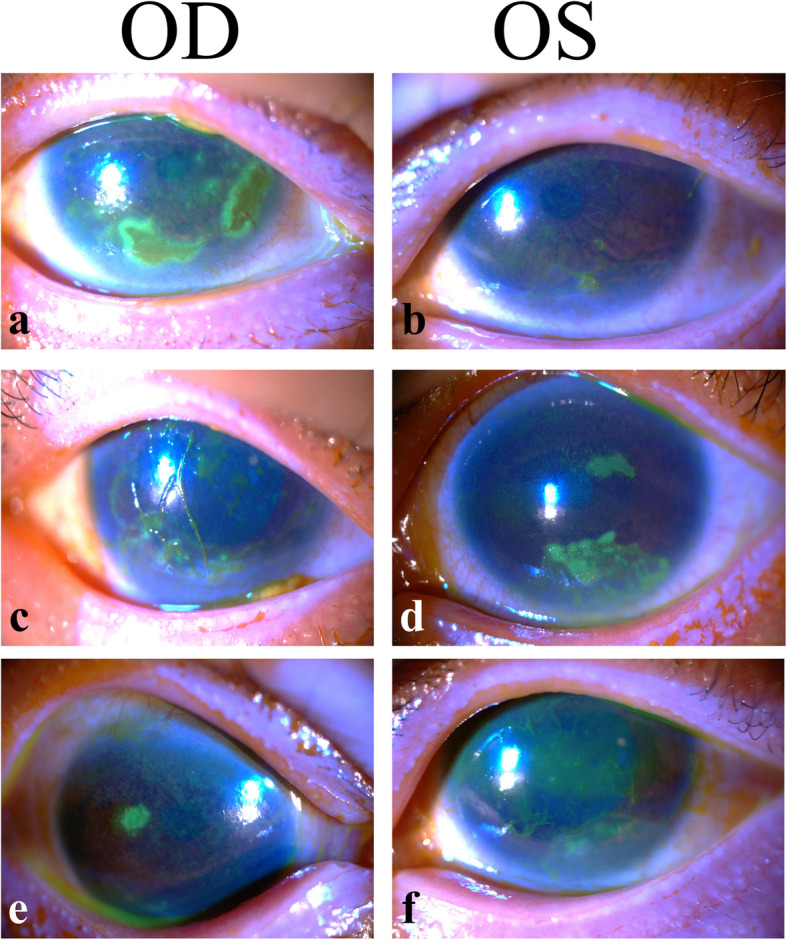
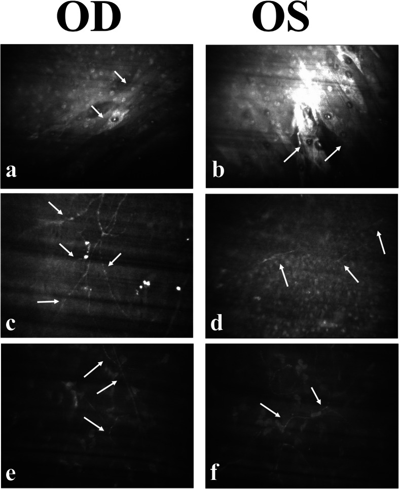
Case presentation: A 27-year-old woman was referred to our hospital because of intermittent blurred vision and recalcitrant ocular surface problems in both eyes for many years. She has a history of autoimmune polyglandular syndrome type 1 (APS-1), which includes hypothyroidism, hypoparathyroidism, hypoadrenalism, and hypogonadotropic hypogonadism. In vivo confocal microscopy clearly demonstrated significant degeneration of the sub-basal nerve plexus and stromal nerve bundles in her corneas bilaterally. She was diagnosed with severe NK and ocular surface disease caused by dry eye. Treatment included the application of therapeutic soft contact lenses and punctual occlusion; however, both treatments had a limited effect.
Conclusion: Patients with APS-1 may have ocular surface disease and severe damage to corneal nerves. Regular follow-up and treatment focusing on the regeneration of corneal nerves is particularly important in these patients.
Keywords: Autoimmune polyglandular syndrome type 1 (APS-1); Case report; In vivo confocal microscopy; Neurotrophic keratitis.
Conflict of interest statement
The authors declare that they have no competing interests.
Figures
Similar articles
-
SARS-CoV-2-induced adrenal crisis in a patient with autoimmune polyglandular syndrome type 1: case report.Folia Med (Plovdiv). 2023 Apr 30;65(2):305-310. doi: 10.3897/folmed.65.e76245. Folia Med (Plovdiv). 2023. PMID: 37144317
-
[Atypical clinical presentation of autoimmune polyglandular syndrome type 4].Przegl Lek. 2011;68(6):339-41. Przegl Lek. 2011. PMID: 22039674 Polish.
-
A Case of Critical Lower-Limb Ischemia in a 29-Year-Old Man with Autoimmune Polyglandular Syndrome Type 1 (APS-1).Am J Case Rep. 2020 Oct 17;21:e924705. doi: 10.12659/AJCR.924705. Am J Case Rep. 2020. PMID: 33068390 Free PMC article.
-
Contact lens induced bacterial keratitis in LCD II: Management and multimodal imaging: a case report and review of literature.Eur J Ophthalmol. 2021 Sep;31(5):2313-2318. doi: 10.1177/1120672120968724. Epub 2020 Oct 30. Eur J Ophthalmol. 2021. PMID: 33124478 Review.
-
[Clinical picture and diagnosis of neurotrophic keratopathy].Ophthalmologe. 2019 Feb;116(2):120-126. doi: 10.1007/s00347-018-0822-x. Ophthalmologe. 2019. PMID: 30535856 Review. German.
Cited by
-
Ocular features of autoimmune polyendocrinopathy candidiasis ectodermal dystrophy.BMJ Case Rep. 2023 Jul 12;16(7):e252672. doi: 10.1136/bcr-2022-252672. BMJ Case Rep. 2023. PMID: 37437958 Free PMC article.
-
Neurotrophic Keratopathy in Systemic Diseases: A Case Series on Patients Treated With rh-NGF.Front Med (Lausanne). 2022 May 30;9:920688. doi: 10.3389/fmed.2022.920688. eCollection 2022. Front Med (Lausanne). 2022. PMID: 35707524 Free PMC article.
References
Publication types
MeSH terms
LinkOut - more resources
Full Text Sources
Other Literature Sources
Miscellaneous
