Case Reports
. 2021 Jan 7;21(1):16.
doi: 10.1186/s12890-020-01358-6.

Enhancing vigilance for cerebral air embolism after pneumonectomy: a case report

Affiliations
Case Reports

Enhancing vigilance for cerebral air embolism after pneumonectomy: a case report

Yijun Mo et al. BMC Pulm Med. .

Abstract

Background: Vascular air embolism (VAE) is a rare but important complication that has not been paid enough attention to in the medical process such as surgery and anesthesia.

Case presentation: We report for the first time that a 54-year-old male patient with central lung cancer developed severe complications of CAE after right pneumonectomy. After targeted first-aid measures such as assisted breathing, mannitol dehydration and antibiotic treatment, the patient gradually improved. The patient became conscious at discharge after 25 days of treatment but left limb was left with nerve injury symptoms.

Conclusion: We analyzed the possible causes of CAE in this case, and the findings from this report would be highly useful as a reference to clinicians.

Keywords: Cerebral air embolism; Neurological recovery; Pneumonectomy.


Conflict of interest statement

The authors declare that they have no competing interests.

Figures

Fig. 1
Fig. 1
Chest CT of right central lung cancer. a. Cross-sectional, lung window showed right central lung cancer. b. Mediastinal window, tumor invaded right main bronchus and right pulmonary artery trunk. c d. Frontal plane, right central lung cancer invade right main bronchus and right pulmonary artery trunk
Fig. 2
Fig. 2
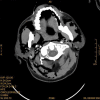
A large amount of gas is present in the interstitial space of the neck
Fig. 3
Fig. 3
CTA results showed that there were no abnormalities such as embolism in the main cerebral artery
Fig. 4
Fig. 4
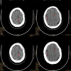
Multiple free air can be seen in the blood vessels of bilateral frontal sulcus as indicated by the arrows
Fig. 5
Fig. 5
Dispersed free air was seen in the vessels of the right occipital lobe and suspicious cerebral infarction lesions were seen
Fig. 6
Fig. 6
Significant reduction in cerebral air embolism 24 h after treatment
Fig. 7
Fig. 7
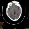
Three days after air embolism, CT showed a patchy low-density shadow in bilateral thalamic basal ganglia (yellow area), temporal lobe (red area) and occipital lobe (black area), which was the cerebral infarction lesion after air embolism

References

    1. Brull SJ, Prielipp RC. Vascular air embolism: a silent hazard to patient safety. J Crit Care. 2017;42:255–263. doi: 10.1016/j.jcrc.2017.08.010. - DOI - PubMed
    1. Schlimp CJ, Bothma PA, Brodbeck AE. Cerebral venous air embolism: what is it and do we know how to deal with it properly? JAMA Neurol. 2014;71:243. doi: 10.1001/jamaneurol.2013.5414. - DOI - PubMed
    1. Hiraki T, Fujiwara H, Sakurai J, Iguchi T, Gobara H, Tajiri N, Mimura H, Kanazawa S. Nonfatal systemic air embolism complicating percutaneous CT-guided transthoracic needle biopsy: four cases from a single institution. Chest. 2007;132(2):684–690. doi: 10.1378/chest.06-3030. - DOI - PubMed
    1. Alper F, Kantarci M, Onbas O, Okur A, Ceviz N. Three-dimensional spiral CT reconstruction in a patient with massive cerebral air embolism. Emerg Radiol. 2004;11:87–88. doi: 10.1007/s10140-004-0358-z. - DOI - PubMed
    1. Schlimp CJ, Loimer T, Rieger M, Lederer W, Schmidts MB. The potential of venous air embolism ascending retrograde to the brain. J Forensic Sci. 2005;50(4):906–909. doi: 10.1520/JFS2005061. - DOI - PubMed

Publication types

MeSH terms