. 2021 Jan 7;21(1):30.
doi: 10.1186/s12935-020-01688-9.

YAP manipulates proliferation via PTEN/AKT/mTOR-mediated autophagy in lung adenocarcinomas

Affiliations

YAP manipulates proliferation via PTEN/AKT/mTOR-mediated autophagy in lung adenocarcinomas

Wei Xu et al. Cancer Cell Int. .

Abstract

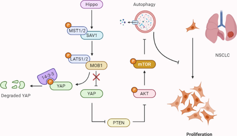
Background: Autophagy is a double-edged sword during the initiation and progression of multiple tumors. The Hippo pathway effector YAP has been proved to be involved in autophagy processes. The present study aimed to investigate how YAP regulates cell proliferation via autophagy in lung adenocarcinomas (LUAD).

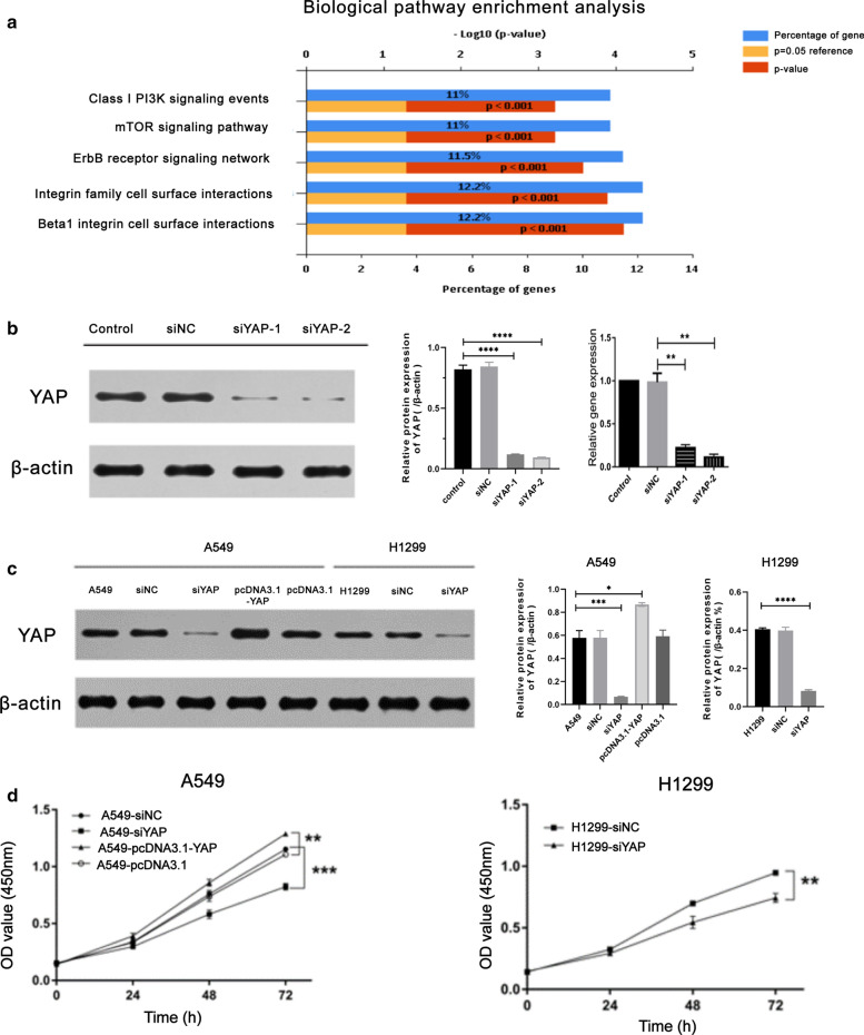
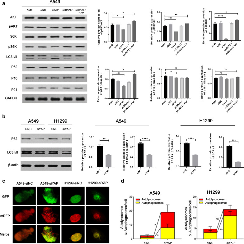
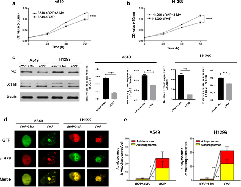
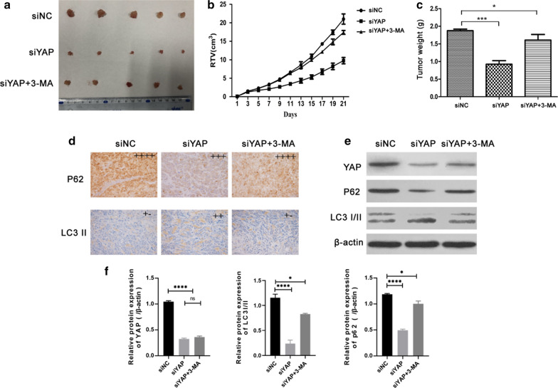
Methods: Data of LUAD chip GSE43458 was obtained from Gene Expression Omnibus (GEO). RT-qPCR and Western blot were performed to assess YAP expression in LUAD cell lines. CCK-8 assay, xenograft tumor model, immunochemistry and GFP-mRFP-LC3 fusion proteins were utilized to evaluate the effect of YAP on autophagy of LUAD cells in vitro and in vivo. Autophagy inhibitor treatment and rescue experiments were carried out to elucidate the mechanism by which YAP manipulates autophagy in LUAD cells.

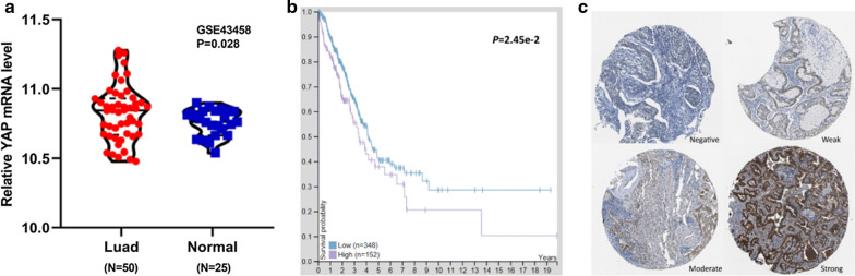
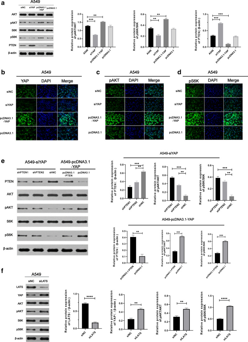
Results: YAP was significantly overexpressed in samples of LUAD patients and its expression level is related to 5-year survival. YAP manipulated the proliferation and autophagy in A549 and H1299 LUAD cells. YAP could induce activation of Akt/mTOR signaling pathway via suppressing PTEN in a Hippo-pathway-dependent manner. 3-Methyladenine impeded autophagy flux and promoted the proliferation in vitro and in vivo.

Conclusions: Hippo pathway critical transcriptional coactivators YAP manipulates the proliferation of lung adenocarcinoma, which is regulated by PTEN/AKT/mTOR autophagic signaling.

Keywords: Autophagy; LUADs; PTEN/AKT/mTOR; YAP.


Conflict of interest statement

The authors declare that there are no conflicts of interests.

Figures

Fig. 1
Fig. 1
YAP is highly expressed in LUAD tissues and predicts poor prognosis. a. The expression of YAP in 50 tissues of LUAD and 25 tissues of normal lung tissues was determined of the GSE43458 at GEO database. b. Kaplan–Meier survival curves and log-rank tests were used to assess the relationship between YAP levels and overall survival time of LUAD patients. The median of YAP expression levels in LUAD tissues was taken as cutoff. c. YAP levels of in four LUAD tissues The Human Protein Atlas: Intensity (negative, weak, moderate, strong) https://www.proteinatlas.org/ENSG00000137693-YAP1/pathology/lung+cancer#imid_19107997
Fig. 2
Fig. 2
YAP targeted siRNA suppresses YAP expression in A549 and H1299 cells with reducing cell proliferation. a The result of biological pathway enrichment analysis was shown with respect to YAP. b siYAP-1/2 was constructed and significantly inhibited the expression of YAP in A549 cells, which detected by qPCR and Western blot. c The protein expression of YAP in A549 transfected with siYAP or pcDNA3.1-YAP, and H1299 with siYAP transfection were analysed by Western blot, respectively. d CCK-8 assay was utilized to analyze cell proliferation of A549 cells with siYAP or pcDNA3.1-YAP, and H1299 with siYAP transfection. Data represent the Mean ± SD on three independent experiments (one-way ANOVA, **P < 0.01, ***P < 0.001)
Fig. 3
Fig. 3
Autophagy in A549 and H1299 cell manipulated by YAP gene. a Analysis of autophagy-related protein expression in A549 cell manipulated by YAP1 gene. Cells were analyzed by immunoblotting with indicated antibodies. b Autophagy substrate proteins p62 and autophagy marker proteins LC3I/II were detected in A549/H1299-siYAP cellular extracts by Western blot. c Immunofluorescence analysis of autolysosomes puncta in A549 and H1299 cells transfected with siYAP. Confocal microscopy images showing cellular localization of autophagic dots in A549/H1299-siYAP cells. The GFP stain (green puncta) was on behalf of the initial process of autophagy and the mRFP (red puncta) indicated the late process of autophagy. Magnification is × 400. d Quantification of autophagy activity was analyzed from 10 random visual fields for each group. At least three different visual fields containing at least 20 cells were counted for each condition and shown in the graph. Data were shown as the Mean ± SD. Statistical analysis was calculated by one-way ANOVA. Scale bars = 25 µm (*P < 0.05, **P < 0.01)
Fig. 4
Fig. 4
YAP induced activation of Akt/mTOR signaling pathway via suppressing PTEN in a Hippo-pathway-dependent manner. a. Following transfected with siYAP or pcDNA3.1-YAP in A549, Western blot measured the level of proteins associated with Akt/mTOR signaling pathway. bd Representative immunofluorescence of A549 cells transfected with siNC, siYAP, pcDNA3.1 or pcDNA3.1-YAP was observed by Inversed Fluorescent Microscope. Fluorescent staining of YAP, pAKT and pS6K was shown as green stain and fluorescence intensity indicated the relative amount in the cells. Magnification is × 200. e Western blot showed the expressive levels of AKT, pAKT, S6K, pS6K, and PTEN after transfection with or without shPTEN in A549-siYAP, with or without pcDNA3.1-PTEN in A549-pcDNA3.1-YAP, respectively. f Western blot showed the expressive levels of AKT, pAKT, S6K, pS6K, and YAP after knockdown LATS, which being a upstream of YAP and a core factor in Hippo pathway
Fig. 5
Fig. 5
The transformation of autophagy in A549/H1299-siYAP after treated with 3-MA. a, b Theproliferationability of A549/H1299-siYAP and A549/H1299-siYAP+3-MA was determined by CCK-8 assay. c A549/H1299-siYAP cells treated with 3-MA were lysated. Autophagy substrate proteins p62 and the conversion of LC3B-I to LC3B-II were detected by the Western blot. de Confocal microscopy images showing cellular localization of autophagic dots in A549/H1299-siYAP or A549/H1299-siYAP + 3MA cells. The GFP stain (green puncta) was on behalf of the initial process of autophagy and the mRFP (red puncta) indicated the late process of autophagy. Magnification is × 400. Quantification of autophagy activity was analyzed from 10 random visual fields for each group and shown in the graph. Data were shown as the Mean ± SD (one-way analysis, *P < 0.05, **P < 0.01, ***P < 0.001)
Fig. 6
Fig. 6
Knockdown of YAP inhibited tumor growth through autophagy in vivo. a Image of tumor size in A549 cells tumor xenograft treated with control, siYAP and siYAP + 3MA. b, c Represented figure indicated the tumor growth after intraperitoneal injection with or without 3MA. d  Immune staining indicated the expression of p62 and LC3 II, scale bar = 200 µm. ef Western blot showed the expressive levels of YAP, p62, and LC3I/II in tumor tissues treated with or without siYAP and 3MA. Data were shown as the Mean ± SD (one-way analysis, *P < 0.05, **P < 0.01, ***P < 0.001)
Fig. 7
Fig. 7
The relationship between YAP and PTEN/AKT/mTOR-mediated autophagy. When the Hippo pathway is revitalite, the activation of core Hippo pathway kinases (MST1/2, SAV1, MOB1A/B or LATS1/2) are ON, and YAP is phosphorylated and cytoplasmically retained by 14-3-3 for degradation. Above process enhancing autophagy by inhibiting AKT/mTOR pathway via elevating PTEN activation, the increase in autophagy further suppressing the non-small cell lung cancer cells proliferation. Once Hippo pathway is inactivated, YAP overexpression inhibits the activity of PTEN, which blocks the AKT/mTOR-mediated autophagy signaling pathway, promoting the proliferation of non-small cell lung cancer cells. Thus, Hippo pathway critical transcriptional coactivators YAP manipulates the proliferation of lung adenocarcinoma, which is regulated by PTEN/AKT/mTOR autophagic signaling

References

    1. Zhao B, Tumaneng K, Guan KL. The Hippo pathway in organ size control, tissue regeneration and stem cell self-renewal. Nat Cell Biol. 2011;13(8):877–83. doi: 10.1038/ncb2303. - DOI - PMC - PubMed
    1. Yu FX, Zhao B, Guan KL. Hippo Pathway in Organ Size Control, Tissue Homeostasis, and Cancer. Cell. 2015;163(4):811–28. doi: 10.1016/j.cell.2015.10.044. - DOI - PMC - PubMed
    1. Zhao B, Wei X, Li W, Udan RS, Yang Q, Kim J, et al. Inactivation of YAP oncoprotein by the Hippo pathway is involved in cell contact inhibition and tissue growth control. Genes Dev. 2007;21(21):2747–61. doi: 10.1101/gad.1602907. - DOI - PMC - PubMed
    1. Zhao B, Li L, Tumaneng K, Wang CY, Guan KL. A coordinated phosphorylation by Lats and CK1 regulates YAP stability through SCF(beta-TRCP) Genes Dev. 2010;24(1):72–85. doi: 10.1101/gad.1843810. - DOI - PMC - PubMed
    1. Meng Z, Moroishi T, Mottier-Pavie V, Plouffe SW, Hansen CG, Hong AW, et al. MAP4K family kinases act in parallel to MST1/2 to activate LATS1/2 in the Hippo pathway. Nat Commun. 2015;6:8357. doi: 10.1038/ncomms9357. - DOI - PMC - PubMed