

microRNA-27b shuttled by mesenchymal stem cell-derived exosomes prevents sepsis by targeting JMJD3 and downregulating NF-κB signaling pathway
- PMID: 33413595
- PMCID: PMC7791667
- DOI: 10.1186/s13287-020-02068-w
microRNA-27b shuttled by mesenchymal stem cell-derived exosomes prevents sepsis by targeting JMJD3 and downregulating NF-κB signaling pathway
Abstract
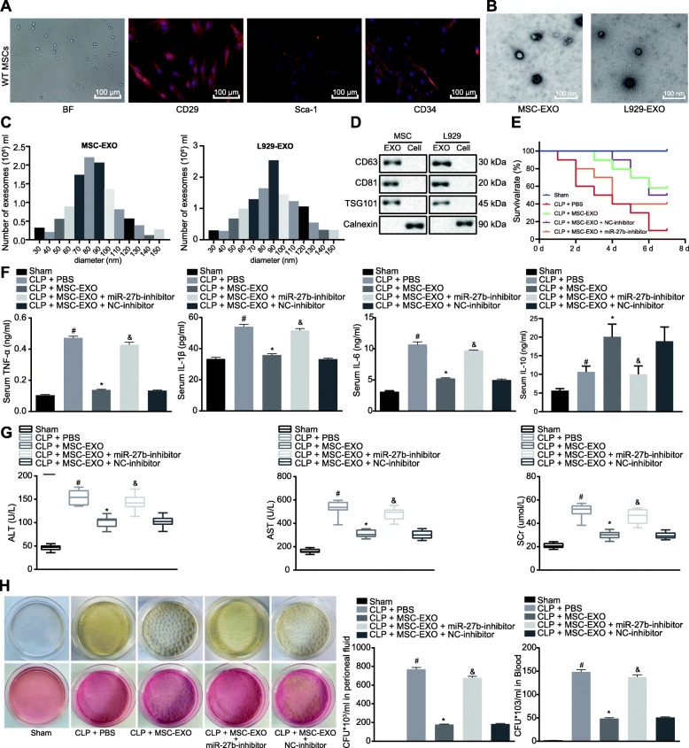
Background: Exosomal microRNAs (miRs) derived from mesenchymal stem cells (MSCs) have been shown to play roles in the pathophysiological processes of sepsis. Moreover, miR-27b is highly enriched in MSC-derived exosomes. Herein, we aimed to investigate the potential role and downstream molecular mechanism of exosomal miR-27b in sepsis.
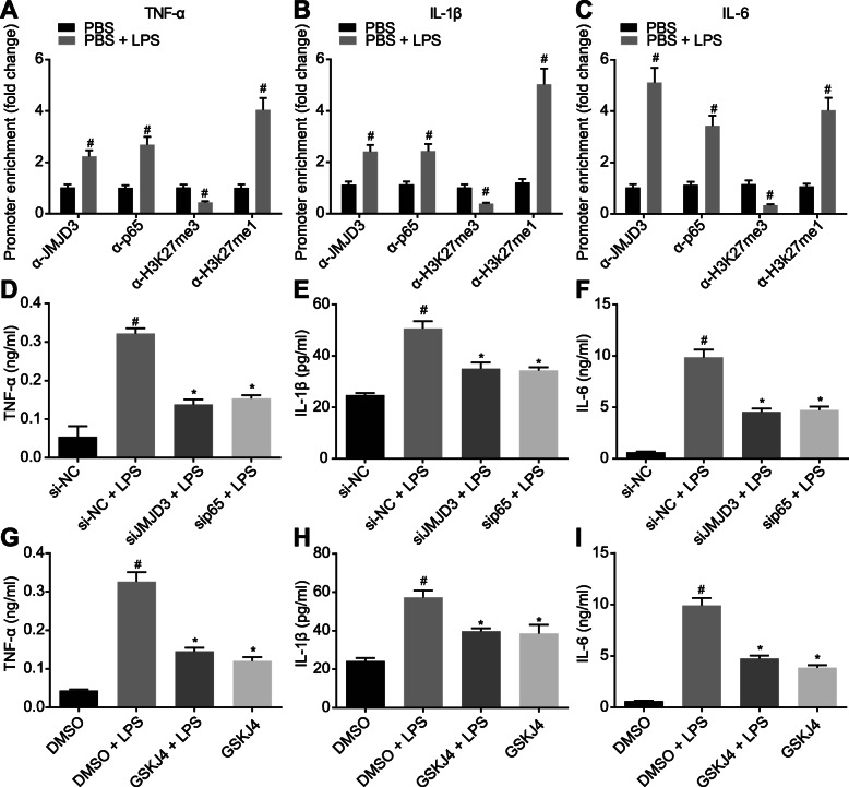
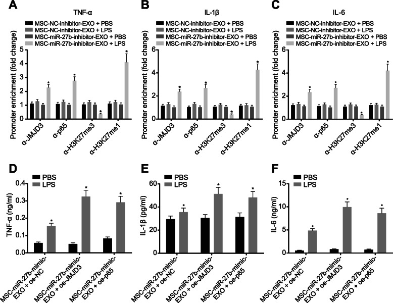
Methods: Inflammation was induced in bone marrow-derived macrophages (BMDMs) by lipopolysaccharide (LPS), and mice were made septic by cecal ligation and puncture (CLP). The expression pattern of miR-27b in MSC-derived exosomes was characterized using RT-qPCR, and its downstream gene was predicted by in silico analysis. The binding affinity between miR-27b, Jumonji D3 (JMJD3), or nuclear factor κB (NF-κB) was characterized to identify the underlying mechanism. We induced miR-27b overexpression or downregulation, along with silencing of JMJD3 or NF-κB to examine their effects on sepsis. The production of pro-inflammatory cytokines TNF-α, IL-1β, and IL-6 was detected by ELISA.
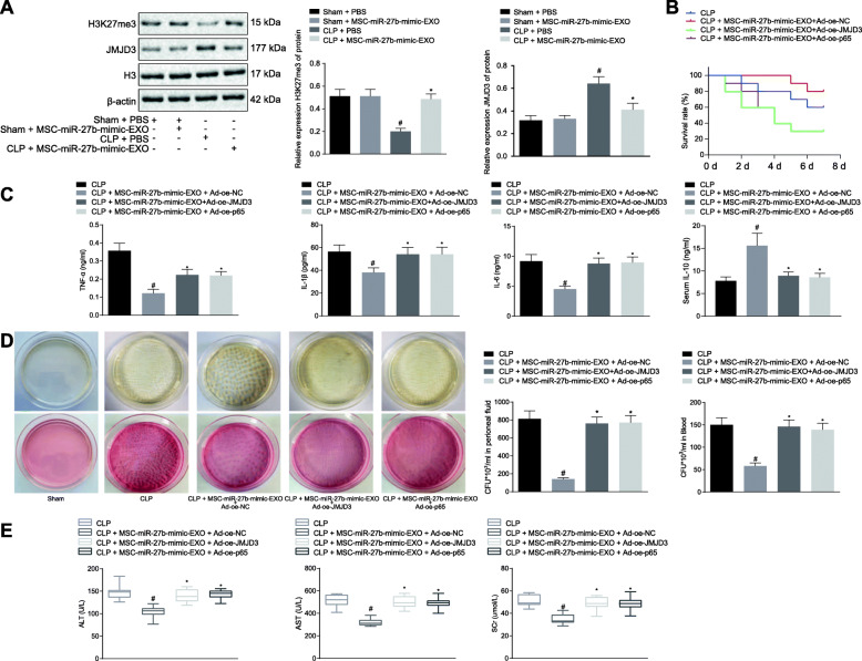
Results: miR-27b was highly expressed in MSC-derived exosomes. Mechanistic investigations showed that miR-27b targeted JMJD3. miR-27b decreased expression of pro-inflammatory genes by inhibiting the recruitment of JMJD3 and NF-κB at gene promoter region. Through this, MSC-derived exosomal miR-27b diminished production of pro-inflammatory cytokines in LPS-treated BMDMs and septic mice, which could be rescued by upregulation of JMJD3 and NF-κB. Besides, in vitro findings were reproduced by in vivo findings.
Conclusion: These data demonstrated that exosomal miR-27b derived from MSCs inhibited the development of sepsis by downregulating JMJD3 and inactivating the NF-κB signaling pathway.
Keywords: Exosome; Jumonji D3; Mesenchymal stem cells; MicroRNA-27b; Nuclear factor κB/p65; Sepsis.
Conflict of interest statement
The authors declare that they have no competing interests.
Figures


Similar articles
-
Human bone mesenchymal stem cells-derived exosomal miRNA-361-5p alleviates osteoarthritis by downregulating DDX20 and inactivating the NF-κB signaling pathway.Bioorg Chem. 2021 Aug;113:104978. doi: 10.1016/j.bioorg.2021.104978. Epub 2021 May 27. Bioorg Chem. 2021. PMID: 34052737
-
Exosomal Micro-RNA-96 Derived From Bone Marrow Mesenchymal Stem Cells Inhibits Doxorubicin-Induced Myocardial Toxicity by Inhibiting the Rac1/Nuclear Factor-κB Signaling Pathway.J Am Heart Assoc. 2021 Sep 7;10(17):e020589. doi: 10.1161/JAHA.120.020589. Epub 2021 Aug 28. J Am Heart Assoc. 2021. PMID: 34459233 Free PMC article.
-
GSKJ4 Protects Mice Against Early Sepsis via Reducing Proinflammatory Factors and Up-Regulating MiR-146a.Front Immunol. 2018 Oct 2;9:2272. doi: 10.3389/fimmu.2018.02272. eCollection 2018. Front Immunol. 2018. PMID: 30337925 Free PMC article.
-
Role of Exosomal miR-223 in Chronic Skeletal Muscle Inflammation.Orthop Surg. 2022 Apr;14(4):644-651. doi: 10.1111/os.13232. Epub 2022 Mar 16. Orthop Surg. 2022. PMID: 35293669 Free PMC article. Review.
-
Exosomes and microRNAs as mediators of the exercise.Eur J Med Res. 2025 Jan 19;30(1):38. doi: 10.1186/s40001-025-02273-4. Eur J Med Res. 2025. PMID: 39828711 Free PMC article. Review.
Cited by
-
Advances in Mesenchymal stem cells regulating macrophage polarization and treatment of sepsis-induced liver injury.Front Immunol. 2023 Oct 25;14:1238972. doi: 10.3389/fimmu.2023.1238972. eCollection 2023. Front Immunol. 2023. PMID: 37954578 Free PMC article. Review.
-
Global Trends and Prospects Regarding Exosomes in Cancer Immunology Research Over the Past 10 Years.Technol Cancer Res Treat. 2023 Jan-Dec;22:15330338231199892. doi: 10.1177/15330338231199892. Technol Cancer Res Treat. 2023. PMID: 37990510 Free PMC article. Review.
-
Protective properties of extracellular vesicles in sepsis models: a systematic review and meta-analysis of preclinical studies.J Transl Med. 2023 Apr 17;21(1):262. doi: 10.1186/s12967-023-04121-7. J Transl Med. 2023. PMID: 37069645 Free PMC article.
-
Clinical Prospect of Mesenchymal Stromal/Stem Cell-Derived Extracellular Vesicles in Kidney Disease: Challenges and the Way Forward.Pharmaceutics. 2023 Jul 9;15(7):1911. doi: 10.3390/pharmaceutics15071911. Pharmaceutics. 2023. PMID: 37514097 Free PMC article. Review.
-
Regulatory Role of Non-Coding RNAs on Immune Responses During Sepsis.Front Immunol. 2021 Dec 9;12:798713. doi: 10.3389/fimmu.2021.798713. eCollection 2021. Front Immunol. 2021. PMID: 34956235 Free PMC article. Review.
References
Publication types
MeSH terms
Substances
LinkOut - more resources
Full Text Sources
Other Literature Sources
Medical
Miscellaneous
