

Iodine-related attenuation in contrast-enhanced dual-energy computed tomography in small-sized solid-type lung cancers is associated with the postoperative prognosis
- PMID: 33413669
- PMCID: PMC7791656
- DOI: 10.1186/s40644-020-00368-1
Iodine-related attenuation in contrast-enhanced dual-energy computed tomography in small-sized solid-type lung cancers is associated with the postoperative prognosis
Abstract
Background: To investigate the correlation between iodine-related attenuation in contrast-enhanced dual-energy computed tomography (DE-CT) and the postoperative prognosis of surgically resected solid-type small-sized lung cancers.
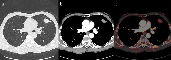
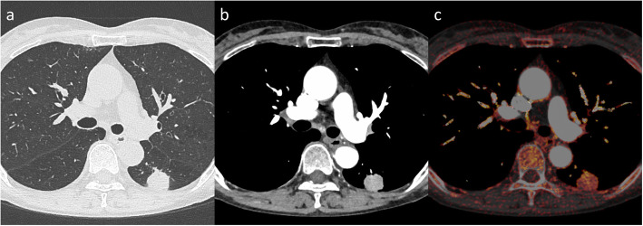
Methods: We retrospectively reviewed the DE-CT findings and postoperative course of solid-type lung cancers ≤3 cm in diameter. After injection of iodinated contrast media, arterial phases were scanned using 140-kVp and 80-kVp tube voltages. Three-dimensional iodine-related attenuation (3D-IRA) of primary tumors at the arterial phase was computed using the "lung nodule" application software. The corrected 3D-IRA normalized to the patient's body weight and contrast medium concentration was then calculated.
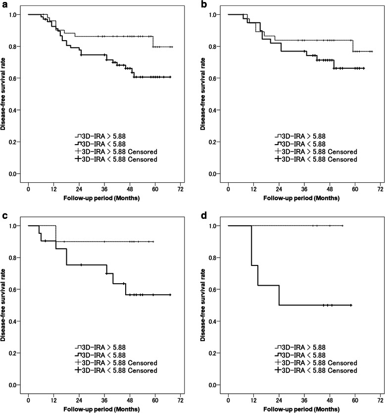
Results: A total of 120 resected solid-type lung cancers ≤3 cm in diameter were selected for analysis (82 males and 38 females; mean age, 67 years). During the observation period (median, 47 months), 32 patients showed postoperative recurrence. Recurrent tumors had significantly lower 3D-IRA and corrected 3D-IRA at early phase compared to non-recurrent tumors (p = 0.046 and p = 0.027, respectively). The area under the receiver operating characteristic curve for postoperative recurrence was 0.624 for the corrected 3D-IRA at early phase (p = 0.025), and the cutoff value was 5.88. Kaplan-Meier curves for disease-free survival indicated that patients showing tumors with 3D-IRA > 5.88 had a significantly better prognosis than those with tumors showing 3D-IRA < 5.88 (p = 0.017).
Conclusions: The 3D-IRA of small-sized solid-type lung cancers on contrast-enhanced DE-CT was significantly associated with postoperative prognosis, and low 3D-IRA tumors showed a higher TNM stage and a significantly poorer prognosis.
Keywords: Contrast enhancement; Dual-energy CT; Functional imaging; Iodine-related attenuation; Lung cancer.
Conflict of interest statement
The authors declare that they have no competing interests.
Figures

Similar articles
-
Evaluation of locoregional invasiveness of small-sized non-small cell lung cancers by enhanced dual-energy computed tomography.Cancer Imaging. 2016 Jul 26;16(1):18. doi: 10.1186/s40644-016-0077-1. Cancer Imaging. 2016. PMID: 27455976 Free PMC article.
-
A comparative analysis of dual-phase dual-energy CT and FDG-PET/CT for the prediction of histopathological invasiveness of non-small cell lung cancer.Eur J Radiol. 2017 Oct;95:186-191. doi: 10.1016/j.ejrad.2017.08.010. Epub 2017 Aug 14. Eur J Radiol. 2017. PMID: 28987666
-
Evaluation of lung cancer by enhanced dual-energy CT: association between three-dimensional iodine concentration and tumour differentiation.Br J Radiol. 2015;88(1055):20150224. doi: 10.1259/bjr.20150224. Epub 2015 Sep 2. Br J Radiol. 2015. PMID: 26329466 Free PMC article.
-
Dual-Energy Computed Tomography Applications in Lung Cancer.Radiol Clin North Am. 2023 Nov;61(6):987-994. doi: 10.1016/j.rcl.2023.06.001. Epub 2023 Aug 4. Radiol Clin North Am. 2023. PMID: 37758365 Review.
-
Review of the application of dual-energy CT combined with radiomics in the diagnosis and analysis of lung cancer.J Appl Clin Med Phys. 2025 Apr;26(4):e70020. doi: 10.1002/acm2.70020. Epub 2025 Feb 17. J Appl Clin Med Phys. 2025. PMID: 39962757 Free PMC article. Review.
Cited by
-
Advances in diagnosis and prediction for aggression of pure solid T1 lung cancer.Precis Clin Med. 2023 Aug 17;6(3):pbad020. doi: 10.1093/pcmedi/pbad020. eCollection 2023 Sep. Precis Clin Med. 2023. PMID: 38025970 Free PMC article.
References
-
- Travis WD, Asamura H, Bankier AA, et al. The IASLC lung Cancer staging project: proposals for coding T categories for subsolid nodules and assessment of tumor size in part-solid tumors in the forthcoming eighth edition of the TNM classification of lung Cancer. J Thorac Oncol. 2016;11(8):1204–1223. doi: 10.1016/j.jtho.2016.03.025. - DOI - PubMed
MeSH terms
Substances
Grants and funding
LinkOut - more resources
Full Text Sources
Other Literature Sources
Medical
