

Dental pulp stem cells overexpressing hepatocyte growth factor facilitate the repair of DSS-induced ulcerative colitis
- PMID: 33413675
- PMCID: PMC7792189
- DOI: 10.1186/s13287-020-02098-4
Dental pulp stem cells overexpressing hepatocyte growth factor facilitate the repair of DSS-induced ulcerative colitis
Abstract
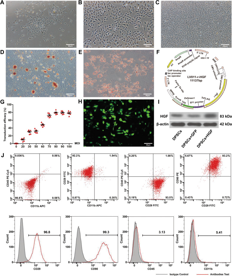
Background: Ulcerative colitis (UC) is a chronic and recurrent disease without satisfactory treatment strategies. Dental pulp stem cell (DPSC) transplantation has been proposed as a potential therapy for UC. This study aimed to investigate the therapeutic effects of the rat hepatocyte growth factor (HGF) gene transduced into DPSCs for UC.
Methods: The therapeutic effects of HGF-DPSCs transplanted intravenously into a rat model of UC induced by 5% dextran sulphate sodium (DSS) were compared with the other treatment groups (LV-HGF group, DPSCs group and GFP-DPSCs group). Immunofluorescence and immunohistochemistry were used to observe the localization and proliferation of HGF-DPSCs at the site of colon injury. The expression levels of inflammatory factors were detected by real-time quantitative PCR (RT-PCR) and western blotting. The oxidative stress markers were detected by ELISA. DAI scores and body weight changes were used to macroscopically evaluate the treatment of rats in each group.
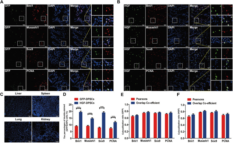
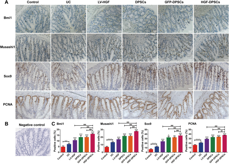
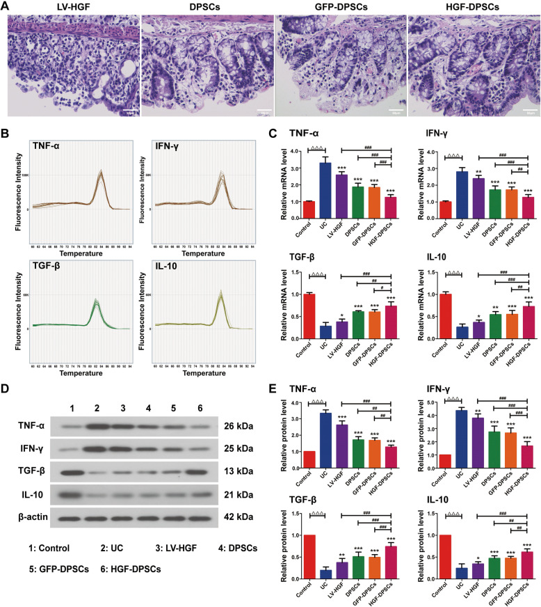
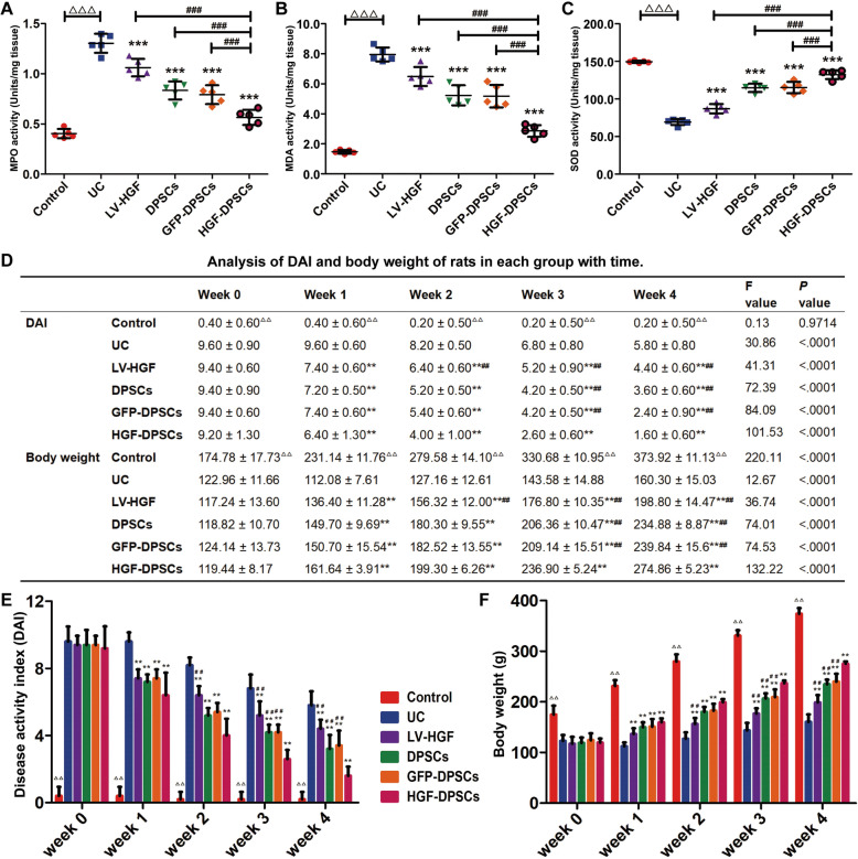
Results: Immunofluorescence and immunohistochemistry assays showed that HGF-DPSCs homed to colon injury sites and colocalized with intestinal stem cell (ISC) markers (Bmi1, Musashi1 and Sox9) and significantly promoted protein expression (Bmi1, Musashi1, Sox9 and PCNA). Anti-inflammatory cytokine (TGF-β and IL-10) expression was the highest in the HGF-DPSCs group compared with the other treatment groups, while the expression of pro-inflammatory cytokines (TNF-α and INF-γ) was the lowest. Additionally, the oxidative stress response results showed that malondialdehyde (MDA) and myeloperoxidase (MPO) expression decreased while superoxide dismutase (SOD) expression increased, especially in the HGF-DPSCs group. The DAI scores showed a downward trend with time in the five treatment groups, whereas body weight increased, and the changes were most prominent in the HGF-DPSCs group.
Conclusions: The study indicated that HGF-DPSCs can alleviate injuries to the intestinal mucosa by transdifferentiating into ISC-like cells, promoting ISC-like cell proliferation, suppressing inflammatory responses and reducing oxidative stress damage, which provides new ideas for the clinical treatment of UC.
Keywords: Dental pulp stem cells; Hepatocyte growth factor; Ulcerative colitis.
Conflict of interest statement
The authors declare that they have no competing interests.
Figures

Similar articles
-
Combinatorial intervention with dental pulp stem cells and sulfasalazine in a rat model of ulcerative colitis.Inflammopharmacology. 2024 Dec;32(6):3863-3879. doi: 10.1007/s10787-024-01532-w. Epub 2024 Jul 29. Inflammopharmacology. 2024. PMID: 39078564 Free PMC article.
-
Canna x generalis L.H. Bailey rhizome extract ameliorates dextran sulfate sodium-induced colitis via modulating intestinal mucosal dysfunction, oxidative stress, inflammation, and TLR4/ NF-ҡB and NLRP3 inflammasome pathways.J Ethnopharmacol. 2021 Apr 6;269:113670. doi: 10.1016/j.jep.2020.113670. Epub 2020 Dec 8. J Ethnopharmacol. 2021. PMID: 33301917
-
Effects of initiating time and dosage of Panax notoginseng on mucosal microvascular injury in experimental colitis.World J Gastroenterol. 2017 Dec 21;23(47):8308-8320. doi: 10.3748/wjg.v23.i47.8308. World J Gastroenterol. 2017. PMID: 29307991 Free PMC article.
-
Pulp stem cells with hepatocyte growth factor overexpression exhibit dual effects in rheumatoid arthritis.Stem Cell Res Ther. 2020 Jun 10;11(1):229. doi: 10.1186/s13287-020-01747-y. Stem Cell Res Ther. 2020. PMID: 32522231 Free PMC article.
-
Overexpression of Hepatocyte Growth Factor in Dental Pulp Stem Cells Ameliorates the Severity of Psoriasis by Reducing Inflammatory Responses.Stem Cells Dev. 2021 Sep 1;30(17):876-889. doi: 10.1089/scd.2021.0129. Epub 2021 Jul 21. Stem Cells Dev. 2021. PMID: 34155928
Cited by
-
Trypsin inhibitor LH011 inhibited DSS-induced mice colitis via alleviating inflammation and oxidative stress.Front Pharmacol. 2022 Sep 27;13:986510. doi: 10.3389/fphar.2022.986510. eCollection 2022. Front Pharmacol. 2022. PMID: 36238566 Free PMC article.
-
Dental Pulp-Derived Stem Cells Preserve Astrocyte Health During Induced Gliosis by Modulating Mitochondrial Activity and Functions.Cell Mol Neurobiol. 2023 Jul;43(5):2105-2127. doi: 10.1007/s10571-022-01291-8. Epub 2022 Oct 6. Cell Mol Neurobiol. 2023. PMID: 36201091 Free PMC article.
-
Therapeutic potential of dental pulp stem cells and their derivatives: Insights from basic research toward clinical applications.World J Stem Cells. 2022 Jul 26;14(7):435-452. doi: 10.4252/wjsc.v14.i7.435. World J Stem Cells. 2022. PMID: 36157522 Free PMC article. Review.
-
Botanicals and Oral Stem Cell Mediated Regeneration: A Paradigm Shift from Artificial to Biological Replacement.Cells. 2022 Sep 7;11(18):2792. doi: 10.3390/cells11182792. Cells. 2022. PMID: 36139367 Free PMC article. Review.
-
Improved therapeutic consistency and efficacy of CD317+ MSCs through stabilizing TSG6 by PTX3.Stem Cell Res Ther. 2024 Mar 27;15(1):92. doi: 10.1186/s13287-024-03706-3. Stem Cell Res Ther. 2024. PMID: 38539221 Free PMC article.
References
-
- Zong S, Pu Y, Li S, Xu B, Zhang Y, Zhang T, Wang B. Beneficial anti-inflammatory effect of paeonol self-microemulsion-loaded colon-specific capsules on experimental ulcerative colitis rats. Artif Cells Nanomed Biotechnol 2018;46:324–335. doi:10.1080/21691401.2017.1423497. - PubMed
Publication types
MeSH terms
Substances
LinkOut - more resources
Full Text Sources
Other Literature Sources
Medical
Research Materials
Miscellaneous
