. 2021 Jan 7;21(1):5.
doi: 10.1186/s40644-020-00371-6.

Assessment of naive indolent lymphoma using whole-body diffusion-weighted imaging and T2-weighted MRI: results of a prospective study in 30 patients

Affiliations

Assessment of naive indolent lymphoma using whole-body diffusion-weighted imaging and T2-weighted MRI: results of a prospective study in 30 patients

Gil-Sun Hong et al. Cancer Imaging. .

Abstract

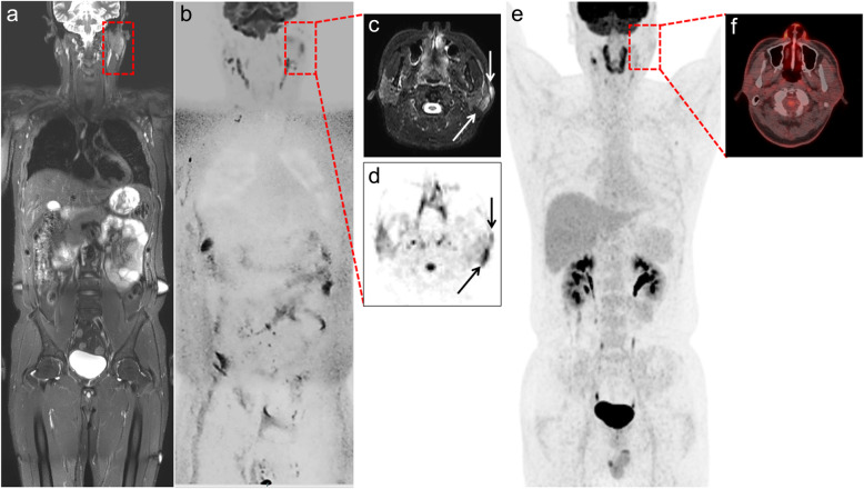
Background: We prospectively evaluated the diagnostic utility of whole-body diffusion-weighted imaging with background body signal suppression and T2-weighted short-tau inversion recovery MRI (WB-DWIBS/STIR) for the pretherapeutic staging of indolent lymphoma in 30 patients.

Methods: This prospective study included 30 treatment-naive patients with indolent lymphomas who underwent WB-DWIBS/STIR and conventional imaging workup plus biopsy. The pretherapeutic staging agreement, sensitivity, and specificity of WB-DWIBS/STIR were investigated with reference to the multimodality and multidisciplinary consensus review for nodal and extranodal lesions excluding bone marrow.

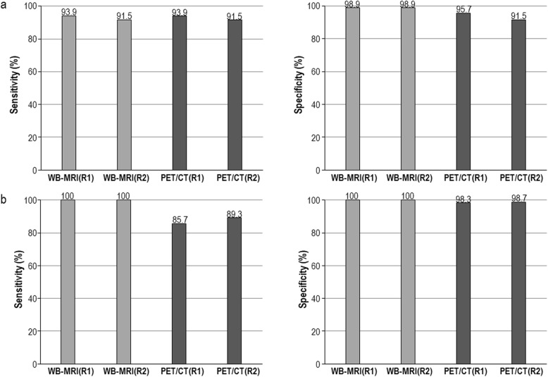
Results: In the pretherapeutic staging, WB-DWIBS/STIR showed very good agreement (κ = 0.96; confidence interval [CI], 0.88-1.00), high sensitivity (93.4-95.1%), and high specificity (99.0-99.4%) for the whole-body regions. These results were similar to those of 18F-FDG-PET/CT, except for the sensitivity for extranodal lesions. For extranodal lesions, WB-DWIBS/STIR showed higher sensitivity compared to 18F-FDG-PET/CT for the whole-body regions (94.9-96.8% vs. 79.6-86.3%, P = 0.058).

Conclusion: WB-DWIBS/STIR is an effective modality for the pretherapeutic staging of indolent lymphoma, and it has benefits when evaluating extranodal lesions, compared with 18F-FDG-PET/CT.

Keywords: Diffusion-weighted imaging with background body signal suppression; Indolent lymphoma; T2-weighted short-tau inversion recovery MRI; Whole-body magnetic resonance imaging.


Conflict of interest statement

The authors declare that they have nothing to disclose.

Figures

Fig. 1
Fig. 1
A 43-year-old man with MALToma of pretherapeutic stage IV. a Coronal whole-body T2-STIR, b DWIBS, c axial T2-STIR, d and axial DWIBS show lymphoma involvement in the left parotid gland and overlying skin (dashed square and arrows). e Coronal whole-body 18F-FDG-PET/CT MIP and (f) 18F-FDG-PET/CT axial fusion images do not show 18F-FDG uptake in the corresponding area. DWIBS = diffusion-weighted imaging with background body signal suppression, T2-STIR = T2-weighted short-tau inversion recovery, 18F-FDG-PET/CT = 18F-fluorodeoxyglucose-positron emission tomography-computed tomography, MIP = maximum intensity projection
Fig. 2
Fig. 2
A 59-year-old man with MALToma of pretherapeutic stage IV. a Coronal whole-body T2-STIR and b DWIBS show lymphoma involvement of the stomach (dashed square) and lymph nodes in the neck (arrow). Magnified DWIBS images show high signal intensity along the greater curvature of the gastric body. c Coronal whole-body 18F-FDG-PET/CT MIP do not show 18F-FDG uptake in the corresponding area of stomach. DWIBS = diffusion-weighted imaging with background body signal suppression, T2-STIR = T2-weighted short-tau inversion recovery, 18F-FDG-PET/CT = 18F-fluorodeoxyglucose-positron emission tomography-computed tomography, MIP = maximum intensity projection
Fig. 3
Fig. 3
Region-based sensitivity and specificity in follicular lymphoma a and MALToma b for WB-DWIBS/STIR and 18F-FDG-PET/CT. WB-MRI = whole-body diffusion-weighted imaging with background body signal suppression and T2-weighted short-tau inversion recovery magnetic resonance imaging, PET/CT = 18F-fluorodeoxyglucose-positron emission tomography-computed tomography, R1 = observer 1, R2 = observer 2

Similar articles

Cited by

References

    1. Galia M, Albano D, Tarella C, Patti C, Sconfienza LM, Mulè A, et al. Whole body magnetic resonance in indolent lymphomas under watchful waiting: the time is now. Eur Radiol. 2018;28:1187–1193. doi: 10.1007/s00330-017-5071-x. - DOI - PubMed
    1. Cheson BD, Fisher RI, Barrington SF, Cavalli F, Schwartz LH, Zucca E, et al. Recommendations for initial evaluation, staging, and response assessment of Hodgkin and non-Hodgkin lymphoma: the Lugano classification. J Clin Oncol. 2014;32:3059–3068. doi: 10.1200/JCO.2013.54.8800. - DOI - PMC - PubMed
    1. Hoffmann M, Wöhrer S, Becherer A, Chott A, Streubel B, Kletter K, et al. 18F-Fluoro-deoxy-glucose positron emission tomography in lymphoma of mucosa-associated lymphoid tissue: histology makes the difference. Ann Oncol. 2006;17:1761–1765. doi: 10.1093/annonc/mdl295. - DOI - PubMed
    1. Perry C, Herishanu Y, Metzer U, Bairey O, Ruchlemer R, Trejo L, et al. Diagnostic accuracy of PET/CT in patients with extranodal marginal zone MALT lymphoma. Eur J Haematol. 2007;79:205–209. doi: 10.1111/j.1600-0609.2007.00895.x. - DOI - PubMed
    1. Radan L, Fischer D, Bar-Shalom R, Dann EJ, Epelbaum R, Haim N, et al. FDG avidity and PET/CT patterns in primary gastric lymphoma. Eur J Nucl Med Mol Imaging. 2008;35:1424–1430. doi: 10.1007/s00259-008-0771-8. - DOI - PubMed

Substances