. 2021 Jan 8;371(6525):pii: eabd4914. doi: 10.1126/science.abd4914.
doi: 10.1126/science.abd4914.

Tubulin glycylation controls axonemal dynein activity, flagellar beat, and male fertility

Affiliations

Tubulin glycylation controls axonemal dynein activity, flagellar beat, and male fertility

Sudarshan Gadadhar et al. Science. .

Abstract

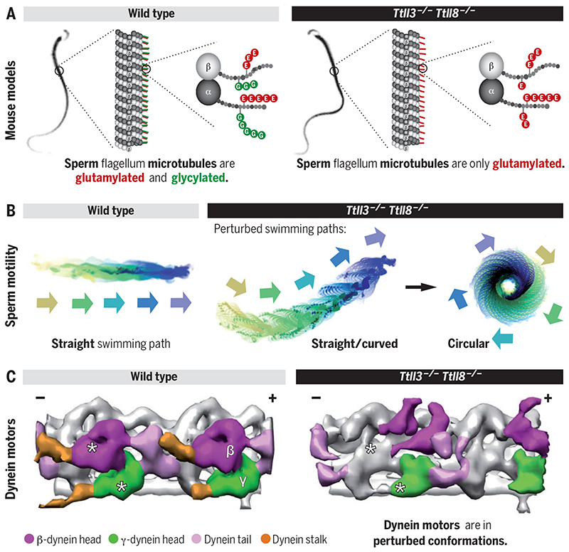
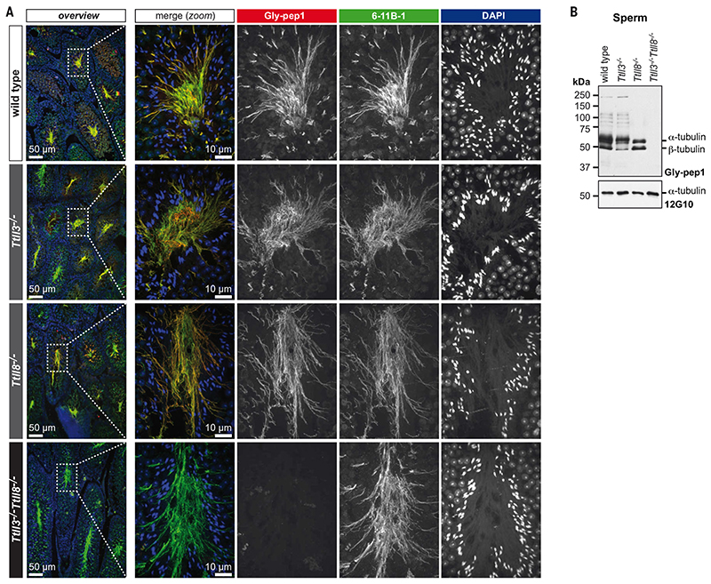
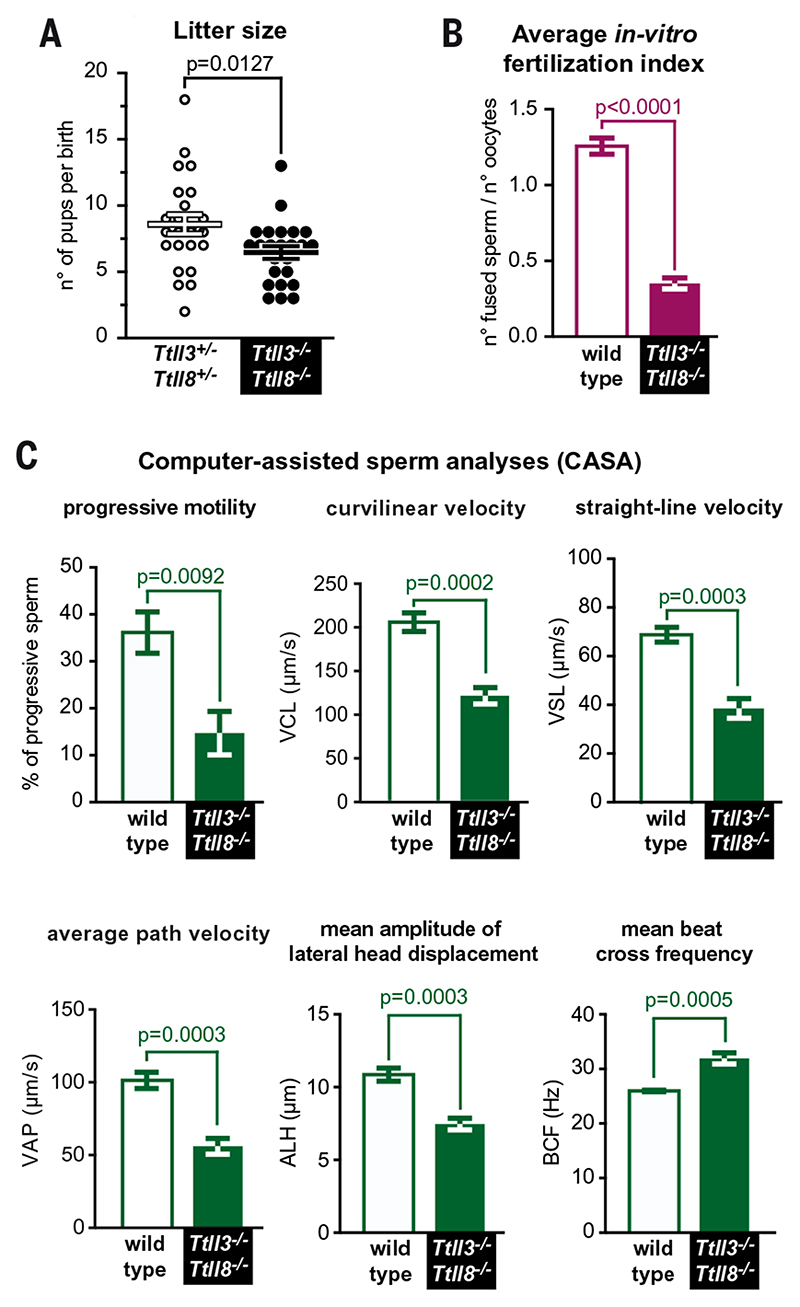
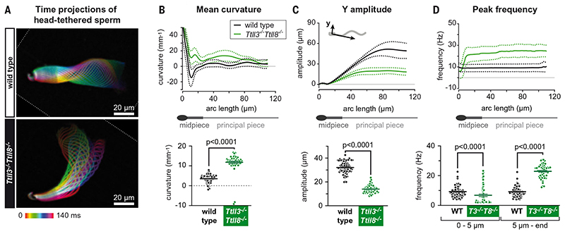
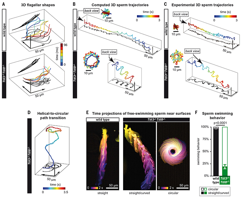
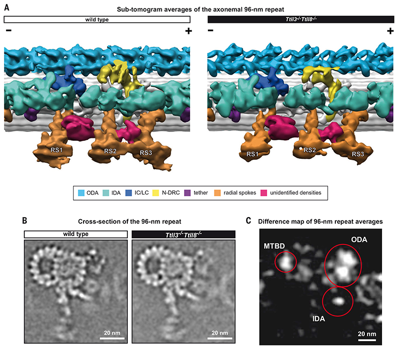
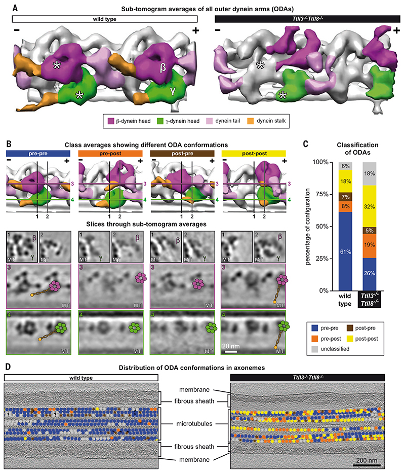
Posttranslational modifications of the microtubule cytoskeleton have emerged as key regulators of cellular functions, and their perturbations have been linked to a growing number of human pathologies. Tubulin glycylation modifies microtubules specifically in cilia and flagella, but its functional and mechanistic roles remain unclear. In this study, we generated a mouse model entirely lacking tubulin glycylation. Male mice were subfertile owing to aberrant beat patterns of their sperm flagella, which impeded the straight swimming of sperm cells. Using cryo-electron tomography, we showed that lack of glycylation caused abnormal conformations of the dynein arms within sperm axonemes, providing the structural basis for the observed dysfunction. Our findings reveal the importance of microtubule glycylation for controlled flagellar beating, directional sperm swimming, and male fertility.


Conflict of interest statement

Competing interests: The authors declare no competing financial interests.

Figures

None
Tubulin glycylation controls sperm motility.
Fig. 1
Fig. 1. Total absence of glycylation in Ttll3 −/− Ttll8 −/− mice.
((A) Paraffin-embedded testes sections stained for tubulin acetylation (6-11B-1; green), glycylation (Gly-pep1; red), and DNA [4′,6-diamidino-2-phenylindole (DAPI)]. Nuclear staining shows normal sperm head morphology and a normal arrangement of sperm heads of different developmental stages in the seminiferous tubules of wild-type, Ttll3 −/−, Ttll8 −/−, and Ttll3 −/− Ttll8 −/− testes. Sperm flagella are present in all genotypes (labeled by 6-11B-1), and are glycylated in wild-type and single-knockout but not in Ttll3 −/− Ttll8 −/− sperm tails. ((B) Immunoblot of sperm samples from wild-type, Ttll3 −/−, Ttll8 −/−, and Ttll3 −/− Ttll8 −/− mice. Note that in single-knockout mice (Ttll3 −/−, Ttll8 −/−), glycylation (Gly-pep1) is only partially abolished, while it is undetectable in Ttll3 −/− Ttll8 −/− sperm. The anti-α-tubulin antibody 12G10 confirmed equal tubulin load.
Fig. 2
Fig. 2. Ttll3 −/− Ttll8 −/− mice show subfertility and reduced sperm motility.
(A) Analysis of litter size. Comparison of heterozygotes (Ttll3 +/− Ttll8 +/−) with Ttll3 /−Ttll8 /− mice shows a reduction in the average number of pups per litter (see fig. S3E for details). (B) In vitro fertilization assay with wild-type oocytes. The average fertilization index of five independent experiments (fig. S4B) shows a strong decrease of Ttll3 −/− Ttll8 −/− sperm fertility. (C) Computer-assisted sperm analyses (CASA) comparing wild-type and Ttll3 −/− Ttll8 −/− sperm. Ttll3 −/− Ttll8 −/− mice show a reduced proportion of progressive sperm. In particular, Ttll3 −/− Ttll8 −/− sperm had reduced velocity parameters: curvilinear velocity (VCL), straight-line velocity (VSL), and average path velocity (VAP), together with reduced amplitude of lateral head displacement (ALH). The beat-cross frequency (BCF), on the other hand, was higher in the Ttll3 −/− Ttll8 −/− sperm.
Fig. 3
Fig. 3. Asymmetric beat of Ttll3 −/− Ttll8 −/− sperm flagella.
(A) Color-coded time projections of dark-field recordings of head-tethered mouse spermatozoa in two dimensions: The depicted color-coded time span (140 ms) corresponds to one beat cycle of wild-type sperm. The flagellar envelope of Ttll3 −/− Ttll8 −/− sperm cells is asymmetrically displaced to the open-hook side of the head, in contrast to wild-type sperm. See also movie S1. (B to D) Multiparameter motility analyses of the flagellar beat using the software SpermQ. In all line graphs, solid lines indicate the time-averaged values, and dotted lines the standard deviation for different arc-length positions along the flagellum. Scatter dot plots show the time- and arc-length-averaged values for individual sperm cells (dots) as well as the mean values ±SEM (bars). (B) Mean flagellar curvature. (C) Amplitude of flagellar beat in the direction perpendicular to the head-midpiece axis. (D) Representation of peak frequencies of the flagellar beat. Scatter plot shows the peak frequencies separately for the first 5 μm and for the rest of the flagellum.
Fig. 4
Fig. 4. Altered swimming behavior of Ttll3 −/− Ttll8 −/− sperm.
(A) 3D flagellar shapes used to compute the 3D swimming path of sperm cells. The shapes were extracted from experiments using tethered sperm (Fig. 3A). The flagellar shapes correspond to about half a beat period, but simulations were based on the complete time series. The z component was assumed to be a smooth arc of constant curvature along the flagellar arc length (κz = 5 × 10⁻³ μm−1; projections shown in black). (B) Computed swimming paths of sperm cells using resistive-force theory. Wild-type spermatozoa are predicted to swim along a twisted ribbon, whereas Ttll3 −/− Ttll8 −/− sperm cells are predicted to swim along a helical path. (C) Representative examples for reconstruction of 3D swimming paths from in-line holographic recordings of freely swimming sperm. Wild-type and Ttll3 −/− Ttll8 −/− sperm swim along a twisted ribbon and a helical path, respectively. Insets [(B) and (C)]: Back view of the path in the direction indicated by the arrow. See also fig. S6 for individual sperm trajectories. (D) 3D holographic trajectory of a Ttll3 −/− Ttll8 −/− sperm showing the transition from a helical to a circular swimming path when reaching the upper wall of the observation chamber. See also movie S3. (E) Color-coded time projections of representative dark-field recordings of freely swimming wild-type and Ttll3 −/− Ttll8 −/− sperm near the glass surface (see also movie S2). (F) Quantification of the swimming patterns observed in (E). The graph represents the mean (±SEM) of the different mice analyzed.
Fig. 5
Fig. 5. The assembly of the axonemal 96-nm repeat is not affected in Ttll3 −/− Ttll8 −/− sperm.
(A) 3D-isosurface rendering of the 96-nm repeats from active wild-type and Ttll3 −/− Ttll8 −/− sperm flagella after subtomogram averaging. All known components of the axonemal 96-nm repeat were identified in both wild type and Ttll3 −/− Ttll8 −/−, indicating that absence of glycylation did not affect their assembly. We further identified densities of structures that are not found in axonemes from other species (crimson-colored). A barrel-shaped structure between radial spokes 1 and 2 (RS1 and RS2) and a density that links the neck of radial spokes 2 and 3 (RS2 and RS3). ODA, outer dynein arm; IDA, inner dynein arm; IC/LC, dynein intermediate chain/light chain; N-DRC, nexin-dynein regulatory complex. (B) Slice through subtomogram averaged wild-type and Ttll3 −/− Ttll8 −/− 96-nm repeats. No evident modifications to the macromolecular assembly of the axoneme were found upon depletion of tubulin glycylation. (C) Difference map of wild-type and Ttll3 −/− Ttll8 −/− 96-nm repeat averages. Areas of the 96-nm repeat that present meaningful structural differences between the two averages are circled in red. These include ODAs, IDAs, and part of the external wall of the B-tubule. MTBD, microtubule-binding domain.
Fig. 6
Fig. 6. Distribution of ODA conformations are perturbed in Ttll3 −/− Ttll8 −/− sperm flagella.
(A) Averages of all ODA subtomograms from wild-type and Ttll3 −/− Ttll8 −/− sperm flagella (from straight axonemal segments) highlight that the structures of the dynein heads (β-heavy chain, magenta; γ-heavy chain, green) are altered in Ttll3 −/− Ttll8 −/− sperm. Asterisks indicate the position of β- and γ-heavy chains in the wild-type structure and the corresponding coordinates in the Ttll3 −/− Ttll8 −/− average. The Ttll3 −/− Ttll8 −/− average shows a shift of both β- and γ-dynein heads toward the MT plus-end (+). (B) Representative class averages of the distinct ODA conformations identified in wild-type and Ttll3 −/− Ttll8 −/− flagella. Isosurface rendering (top panels) and representative orthogonal and longitudinal slices (lower panels) through β- and γ-heavy chains [color-coded as in (A)]. The numbered lines illustrate the slicing planes through the subtomogram averages in the lower panels. The superimposed schematic models show the positions of dynein β- and γ-AAA domains (magenta and green, respectively), the dynein stalks (orange lines), and the MT binding domains (orange dot). Note that pre-pre, pre-post, post-pre, and post-post refer to dynein γ- and β-heavy chain conformations, respectively (pre = pre-powerstroke, post = post-powerstroke). While the position of the stalk is clearly visible in the pre-pre and post-post conformations, as highlighted by the schematics, the stalks were not easily identified in the pre-post and post-pre conformations, probably because of their unstable conformation. (C) Incidence of the distinct ODA conformations in wild-type (n = 2691 particles) and Ttll3 −/− Ttll8 −/− (n = 3656 particles) axonemes. Ttll3 −/− Ttll8 −/− flagella show a particularly increased percentage of pre-post and post-post powerstroke conformations. (D) Distribution of the different ODAs conformations visualized in wild-type and Ttll3 −/− Ttll8 −/− axonemes shows the absence of a clear pattern of distribution in Ttll3 −/− Ttll8 −/− sperm [color coding of dots as in (B) and (C)]. The distribution in Ttll3 −/− Ttll8 −/− indicates a reduced ability of ODAs to coordinate their activity states.

Comment in

  • Sperm going in circles.
    Strzyz P. Strzyz P. Nat Rev Mol Cell Biol. 2021 Apr;22(4):242-243. doi: 10.1038/s41580-021-00345-x. Nat Rev Mol Cell Biol. 2021. PMID: 33558683 No abstract available.

Similar articles

Cited by

References

    1. Janke C, Magiera MM. The tubulin code and its role in controlling microtubule properties and functions. Nat Rev Mol Cell Biol. 2020;21:307–326. doi: 10.1038/s41580-020-0214-3. - DOI - PubMed
    1. Bré MH, et al. Axonemal tubulin polyglycylation probed with two monoclonal antibodies: Widespread evolutionary distribution, appearance during spermatozoan maturation and possible function in motility. J Cell Sci. 1996;109:727–738. - PubMed
    1. Rogowski K, et al. Evolutionary divergence of enzymatic mechanisms for posttranslational polyglycylation. Cell. 2009;137:1076–1087. doi: 10.1016/j.cell.2009.05.020. - DOI - PubMed
    1. Bosch Grau M, et al. Tubulin glycylases and glutamylases have distinct functions in stabilization and motility of ependymal cilia. J Cell Biol. 2013;202:441–451. doi: 10.1083/jcb.201305041. - DOI - PMC - PubMed
    1. Bosch Grau M, et al. Alterations in the balance of tubulin glycylation and glutamylation in photoreceptors leads to retinal degeneration. J Cell Sci. 2017;130:938–949. doi: 10.1242/jcs.199091. - DOI - PubMed

Publication types