. 2021 Jan 8;371(6525):194-200.
doi: 10.1126/science.abc0476.

Structural basis for antibody inhibition of flavivirus NS1-triggered endothelial dysfunction

Affiliations

Structural basis for antibody inhibition of flavivirus NS1-triggered endothelial dysfunction

Scott B Biering et al. Science. .

Abstract

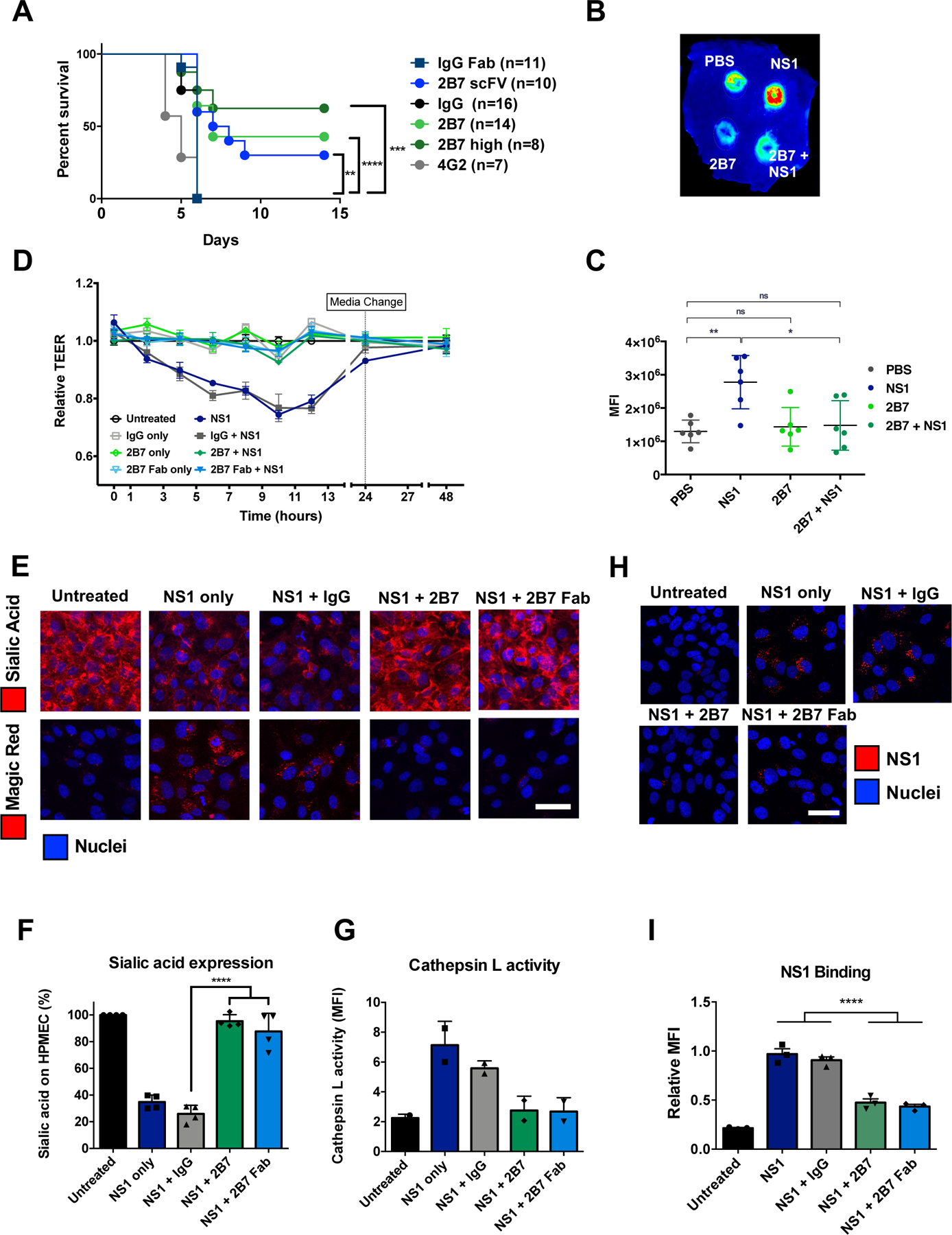
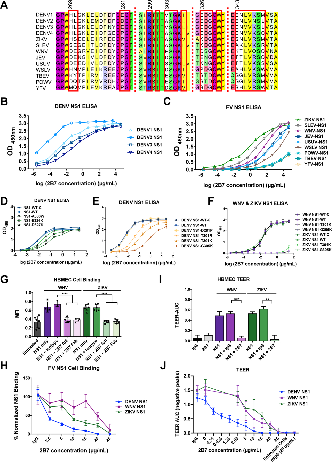
Medically important flaviviruses cause diverse disease pathologies and collectively are responsible for a major global disease burden. A contributing factor to pathogenesis is secreted flavivirus nonstructural protein 1 (NS1). Despite demonstrated protection by NS1-specific antibodies against lethal flavivirus challenge, the structural and mechanistic basis remains unknown. Here, we present three crystal structures of full-length dengue virus NS1 complexed with a flavivirus-cross-reactive, NS1-specific monoclonal antibody, 2B7, at resolutions between 2.89 and 3.96 angstroms. These structures reveal a protective mechanism by which two domains of NS1 are antagonized simultaneously. The NS1 wing domain mediates cell binding, whereas the β-ladder triggers downstream events, both of which are required for dengue, Zika, and West Nile virus NS1-mediated endothelial dysfunction. These observations provide a mechanistic explanation for 2B7 protection against NS1-induced pathology and demonstrate the potential of one antibody to treat infections by multiple flaviviruses.


Conflict of interest statement

Competing interests: Authors declare no competing interests.

Figures

Fig. 1.
Fig. 1.. Anti-NS1 mAb 2B7 is protective against lethal dengue virus infection and NS1-mediated vascular leak and endothelial dysfunction.
(A) Survival curve of Ifnar−/− mice infected with DENV2-D220. Mice were given two 150-μg doses (300 μg for “2B7 high”) of full-length 2B7, a 2B7 single-chain variable fragment (scFv), an anti-E antibody (4G2), or an isotype control antibody the day before and after infection. Numbers in parentheses indicate the number of mice in each group. (B) Localized leak of the tracer molecule, dextran-647, was measured after dorsal intradermal injection of NS1 with or without 2B7, or the indicated controls, into the shaved backs of mice. One representative experiment of n=6 mice is displayed. (C) Quantification of B as mean fluorescence intensity (MFI). (D) TEER assay of HPMEC hyperpermeability after addition of DENV2 NS1 with or without 2B7, or the indicated controls, at the indicated time-points post-NS1 treatment. n=3 biological replicates. (E) Endothelial dysfunction and EGL disruption was monitored using immunofluorescent microscopy 6 hours post-treatment of DENV2 NS1 with or without 2B7, 2B7 Fab, or the indicated controls. Endothelial cell dysfunction (bottom, n=2 biological replicates) was monitored using the cathepsin L-activity reporter molecule, Magic Red, and EGL disruption (top, n=4 biological replicates) was monitored by staining sialic acid on the cell surface. (F) and (G) Quantification of E. (H) DENV2 NS1 binding to HPMEC in the presence of 2B7, 2B7 Fab, or the indicated controls, was monitored by immunofluorescent microscopy 90 minutes post-NS1treatment. n=3 biological replicates. (I) Quantification of H. For all figures, scale bars are 50 μm. n.s., not significant p > 0.05; *p < 0.05; **p < 0.01; ***p < 0.001; ****p < 0.0001 by one-way ANOVA analysis with multiple comparisons.
Fig. 2.
Fig. 2.. Crystal structure of the 2B7 antigen-binding fragment complexed with DENV NS1 reveals binding to β-ladder.
(A) Perpendicular views of a 3.3-Å crystal structure of 2B7 Fab (heavy chain, dark blue; light chain, light blue) and DENV1 NS1 dimer (β-ladder domains, green; β-roll and wing domains, cyan). The combining site is boxed (yellow) in the lower image, right monomer. (B) Detail of the 2B7 scFv and the DENV2 NS1 combining site highlighting the interacting amino acids. The 2B7 backbone is in blue, and NS1 is in green with key side chains shown as sticks. Pan-flavivirus conserved side chains (orange) are at the center of the discontinuous epitope; DENV-conserved side chains (yellow) are at the epitope periphery; hydrogen bonds are shown as dashed lines. (C) DENV2 NS1 epitope for 2B7 scFv (colored by conservation across flaviviruses according to the key and based on the alignment in Fig. 3A), with surfaces outside the epitope in gray. Sites of mutagenesis in Fig. 3 are labeled. (D) 2B7 scFv complementarity-determining regions (CDRs, in tube rendering) for the heavy chain (dark blue) and light chain (light blue) overlaid on the DENV2 NS1 epitope surface. Surfaces of amino acids conserved among the four DENV serotypes but variable in other flaviviruses are purple, other epitope residues are in green; view as in (C).
Fig. 3.
Fig. 3.. 2B7 cross-reacts with multiple flavivirus NS1 proteins.
(A) Alignment of amino acids across the discontinuous 2B7 combining site in NS1 from different flaviviruses. Residues in contact with 2B7 are boxed in red. (B) ELISAs measuring the interaction of 2B7 with NS1 from DENV1–4. Data displayed are at least n=3 biological replicates. (C) ELISAs measuring 2B7 interaction with other flavivirus NS1 proteins (at least n=3 biological replicates). (D) and (E) Same as (C) except for the indicated DENV2 NS1 mutagenized proteins compared to the in-house produced (NS1-WT) or commercially purchased (NS1-WT-C) control proteins. Data displayed are n=3 biological replicates. (F) Same as (C) except for the indicated WNV or ZIKV NS1 mutagenized proteins (n=3 biological replicates). (G) WNV or ZIKV NS1 binding to HBMEC in the presence or absence of 25 g/ml 2B7, and the indicated controls, was measured by immunofluorescent microscopy 90 minutes post-NS1 treatment. Data displayed are at least n=4 biological replicates. (H) Same as (G) but with the indicated concentrations of 2B7 (at least n=3 biological replicates). (I) The effect of WNV or ZIKV NS1 on hyperpermeability of HBMEC was measured using TEER, in the presence or absence of 25 g/ml 2B7, or the indicated controls. AUC is the negative area under the curve (fig. S8C) correlating with a drop in endothelial cell monolayer electrical resistance. Data presented are n=2 biological replicates. (J) Same as (I) but with the indicated concentration of 2B7 (at least n=3 biological replicates). **p < 0.01; ***p < 0.001; ****p < 0.0001 by one-way ANOVA analysis with multiple comparisons. FV, flavivirus.
Fig. 4.
Fig. 4.. Mechanistic insight into 2B7 blockade of NS1-mediated endothelial dysfunction.
(A) 2B7 Fab (blue ribbon) in complex with DENV1 NS1 hexamer above a schematic of the plasma membrane, illustrating 2B7 Fab-mediated steric hinderance of NS1 membrane interaction. The NS1 surface is colored by dimer (light/dark green at right, gray at left and tan in the back), with hydrophobic regions in yellow for all three dimers. The 2B7 Fab is bound to both subunits of the green dimer and occludes cell surface interaction of the yellow wing-domain hydrophobic loop (centered on the WWG motif). (B) Perpendicular views of 2B7 Fab complex with DENV1 NS1 dimer, illustrating Fab interference with membrane interaction of the NS1 hydrophobic face (yellow). The left image shows the dimer hydrophobic face with wing hydrophobic loops at the periphery and central hydrophobic surface of the β-roll domain. This face is inside the hexamer and invisible in (A). (See Movie S1.) The NS1 epitope for 2B7 is in orange. (C) Endothelial hyperpermeability of HPMEC was monitored via TEER for the indicated DENV2 NS1 mutants compared to the commercial NS1-WT (NS1-WT-C) or in-house produced NS1-WT (NS1-WT). Data presented are n=2 biological replicates. (D) Cell binding to HPMEC of NS1 mutants or controls was monitored by immunofluorescent microscopy 90 minutes post-NS1 treatment (n=3 biological replicates). (E) Endothelial dysfunction was monitored using immunofluorescent microscopy and the cathepsin L-activity reporter molecule, Magic Red, 6 hours post-treatment of DENV2 NS1 mutants, or the indicated controls. Data presented are n=2 biological replicates. (F) Quantification of E. n.s., not significant p > 0.05; *p < 0.05; **p < 0.01 by one-way ANOVA analysis with multiple comparisons.

Comment in

References

    1. Bhatt S et al., Nature 496, 504–507 (2013). - PMC - PubMed
    1. Halstead SB, Lancet 370, 1644–1652 (2007). - PubMed
    1. Beatty PR et al., Sci Transl Med 7, 304ra141 (2015). - PubMed
    1. Modhiran N et al., Sci Transl Med 7, 304ra142 (2015). - PubMed
    1. Rothman AL, Nat Rev Immunol 11, 532–543 (2011). - PubMed

Publication types

MeSH terms