

doi: 10.3174/ajnr.A7022.
Epub 2021 Jan 7.
Neuroimaging Offers Low Yield in Children Positive for SARS-CoV-2
Affiliations
- PMID: 33414225
- PMCID: PMC8115373
- DOI: 10.3174/ajnr.A7022
Item in Clipboard
Neuroimaging Offers Low Yield in Children Positive for SARS-CoV-2
AJNR Am J Neuroradiol.
2021 May.
Abstract
The coronavirus disease 2019 (COVID-19) pandemic caused by Severe Acute Respiratory Syndrome coronavirus disease 2 (SARS CoV-2) most commonly presents with respiratory disease, but neurologic complications are being reported. We aimed to investigate the rate of positive neuroimaging findings in children positive for SARS-CoV-2 referred for neuroimaging between March 18 and September 30, 2020. We found that 10% (n = 2) had acute findings. Our results may suggest that in children, neurologic involvement in COVID-19 is rare, neuroimaging has a low yield in diagnosis, and acute neuroimaging should involve careful risk-benefit analysis.
© 2021 by American Journal of Neuroradiology.
Figures
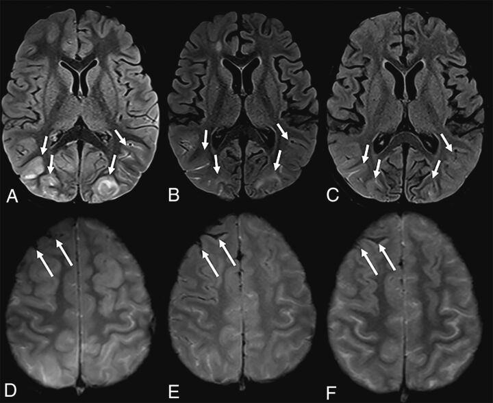
A 7-year-old boy with sickle cell disease who presented with dyspnea and chest pain tested positive for SARS-CoV-2. The patient was unresponsive, having desaturations and being intubated. Brain MR imaging showed T2-FLAIR hyperintensity and cortical edema in the occipital lobes, consistent with posterior reversible encephalopathy syndrome, partially resolving on subsequent imaging (A–C, arrows). Note interval evolution of right frontal subarachnoid hemorrhage (D–F, arrows).
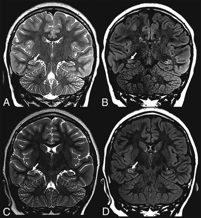
A previously healthy 9-year-old girl who presented with status epilepticus tested positive for SARS-CoV-2. Brain MR imaging showed subtle right hippocampal T2-FLAIR signal alteration with corresponding edema (arrows) on initial (upper row) and follow-up (lower row) imaging.
Similar articles
-
Brain magnetic resonance imaging findings in children with neurological complications of coronavirus disease 2019 (Omicron variant): a multicenter retrospective observational study.Pediatr Radiol. 2024 May;54(6):1012-1021. doi: 10.1007/s00247-024-05908-6. Epub 2024 Mar 28. Pediatr Radiol. 2024. PMID: 38538753
-
Neuroimaging manifestations in children with SARS-CoV-2 infection: a multinational, multicentre collaborative study.Lancet Child Adolesc Health. 2021 Mar;5(3):167-177. doi: 10.1016/S2352-4642(20)30362-X. Epub 2020 Dec 16. Lancet Child Adolesc Health. 2021. PMID: 33338439 Free PMC article.
-
Neuroradiologic manifestations of COVID-19: what the emergency radiologist needs to know.Emerg Radiol. 2020 Dec;27(6):737-745. doi: 10.1007/s10140-020-01840-y. Epub 2020 Aug 21. Emerg Radiol. 2020. PMID: 32822060 Free PMC article. Review.
-
Human and novel coronavirus infections in children: a review.Paediatr Int Child Health. 2021 Feb;41(1):36-55. doi: 10.1080/20469047.2020.1781356. Epub 2020 Jun 25. Paediatr Int Child Health. 2021. PMID: 32584199 Review.
-
Severity of Chest Imaging is Correlated with Risk of Acute Neuroimaging Findings among Patients with COVID-19.AJNR Am J Neuroradiol. 2021 May;42(5):831-837. doi: 10.3174/ajnr.A7032. Epub 2021 Feb 4. AJNR Am J Neuroradiol. 2021. PMID: 33541897 Free PMC article.
Cited by
-
Neurological effects of COVID-19 in infants and children.Dev Med Child Neurol. 2022 Jul;64(7):818-829. doi: 10.1111/dmcn.15185. Epub 2022 Mar 3. Dev Med Child Neurol. 2022. PMID: 35243616 Free PMC article. Review.
-
Pediatric Post-Acute Sequelae of SARS-CoV-2 Infection.Fatigue. 2023;11(2-4):55-65. doi: 10.1080/21641846.2022.2162764. Epub 2023 Jan 1. Fatigue. 2023. PMID: 38044956 Free PMC article.
-
Acute Respiratory Distress Syndrome Associated with Multisystem Inflammatory Syndrome in a Child with Covid-19 and Diabetic Ketoacidosis: A Case Report.Pulm Ther. 2022 Sep;8(3):333-342. doi: 10.1007/s41030-022-00192-x. Epub 2022 May 24. Pulm Ther. 2022. PMID: 35608797 Free PMC article.
-
Brain magnetic resonance imaging findings in children with neurological complications of coronavirus disease 2019 (Omicron variant): a multicenter retrospective observational study.Pediatr Radiol. 2024 May;54(6):1012-1021. doi: 10.1007/s00247-024-05908-6. Epub 2024 Mar 28. Pediatr Radiol. 2024. PMID: 38538753
-
Topographical Distribution of Neuroanatomical Abnormalities Following COVID-19 Invasion : A Systematic Literature Review.Clin Neuroradiol. 2024 Mar;34(1):13-31. doi: 10.1007/s00062-023-01344-5. Epub 2023 Sep 11. Clin Neuroradiol. 2024. PMID: 37697012 Free PMC article.
References
MeSH terms
LinkOut - more resources
Full Text Sources
Other Literature Sources
Medical
Miscellaneous