Regional and Volumetric Parameters for Diffusion-Weighted WHO Grade II and III Glioma Genotyping: A Method Comparison
- PMID: 33414227
- PMCID: PMC7959449
- DOI: 10.3174/ajnr.A6965
Regional and Volumetric Parameters for Diffusion-Weighted WHO Grade II and III Glioma Genotyping: A Method Comparison
Abstract
Background and purpose: Studies consistently report lower ADC values in isocitrate dehydrogenase (IDH) wild-type gliomas than in IDH mutant tumors, but their methods and thresholds vary. This research aimed to compare volumetric and regional ADC measurement techniques for glioma genotyping, with a focus on IDH status prediction.
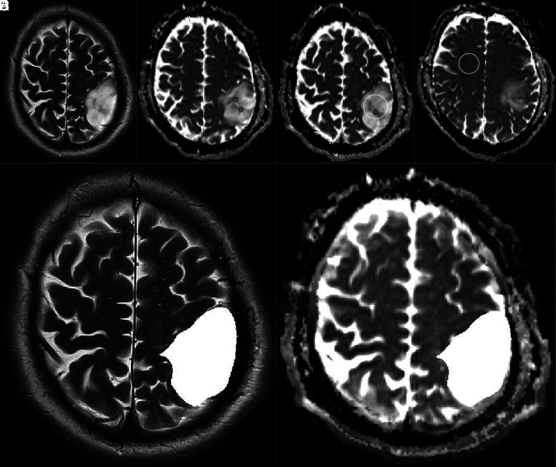
Materials and methods: Treatment-naïve World Health Organization grade II and III gliomas were analyzed by 3 neuroradiologist readers blinded to tissue results. ADC minimum and mean ROIs were defined in tumor and in normal-appearing white matter to calculate normalized values. T2-weighted tumor VOIs were registered to ADC maps with histogram parameters (mean, 2nd and 5th percentiles) extracted. Nonparametric testing (eta2 and ANOVA) was performed to identify associations between ADC metrics and glioma genotypes. Logistic regression was used to probe the ability of VOI and ROI metrics to predict IDH status.
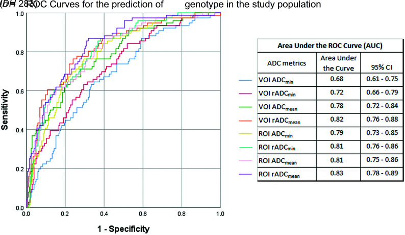
Results: The study included 283 patients with 79 IDH wild-type and 204 IDH mutant gliomas. Across the study population, IDH status was most accurately predicted by ROI mean normalized ADC and VOI mean normalized ADC, with areas under the curve of 0.83 and 0.82, respectively. The results for ROI-based genotyping of nonenhancing and solid-patchy enhancing gliomas were comparable with volumetric parameters (area under the curve = 0.81-0.84). In rim-enhancing, centrally necrotic tumors (n = 23), only volumetric measurements were predictive (0.90).
Conclusions: Regional normalized mean ADC measurements are noninferior to volumetric segmentation for defining solid glioma IDH status. Partially necrotic, rim-enhancing tumors are unsuitable for ROI assessment and may benefit from volumetric ADC quantification.
© 2021 by American Journal of Neuroradiology.
Figures


Similar articles
-
World Health Organization Grade II/III Glioma Molecular Status: Prediction by MRI Morphologic Features and Apparent Diffusion Coefficient.Radiology. 2020 Jul;296(1):111-121. doi: 10.1148/radiol.2020191832. Epub 2020 Apr 21. Radiology. 2020. PMID: 32315266
-
Apparent diffusion coefficient for molecular subtyping of non-gadolinium-enhancing WHO grade II/III glioma: volumetric segmentation versus two-dimensional region of interest analysis.Eur Radiol. 2018 Sep;28(9):3779-3788. doi: 10.1007/s00330-018-5351-0. Epub 2018 Mar 23. Eur Radiol. 2018. PMID: 29572636 Free PMC article.
-
Predicting Genotype and Survival in Glioma Using Standard Clinical MR Imaging Apparent Diffusion Coefficient Images: A Pilot Study from The Cancer Genome Atlas.AJNR Am J Neuroradiol. 2018 Oct;39(10):1814-1820. doi: 10.3174/ajnr.A5794. Epub 2018 Sep 6. AJNR Am J Neuroradiol. 2018. PMID: 30190259 Free PMC article.
-
The T2-FLAIR-mismatch sign as an imaging biomarker for IDH and 1p/19q status in diffuse low-grade gliomas: a systematic review with a Bayesian approach to evaluation of diagnostic test performance.Neurosurg Focus. 2019 Dec 1;47(6):E13. doi: 10.3171/2019.9.FOCUS19660. Neurosurg Focus. 2019. PMID: 31786548
-
Isocitrate dehydrogenase mutation and risk of venous thromboembolism in glioma: A systematic review and meta-analysis.Thromb Res. 2022 Nov;219:14-21. doi: 10.1016/j.thromres.2022.08.029. Epub 2022 Sep 6. Thromb Res. 2022. PMID: 36088710
Cited by
-
Volumetric apparent diffusion coefficient (ADC) histogram analysis of the brain in paediatric patients with hypoxic ischaemic encephalopathy.Pol J Radiol. 2023 Sep 6;88:e399-e406. doi: 10.5114/pjr.2023.131696. eCollection 2023. Pol J Radiol. 2023. PMID: 37808174 Free PMC article.
-
Combining Multi-Shell Diffusion with Conventional MRI Improves Molecular Diagnosis of Diffuse Gliomas with Deep Learning.Cancers (Basel). 2023 Jan 12;15(2):482. doi: 10.3390/cancers15020482. Cancers (Basel). 2023. PMID: 36672430 Free PMC article.
-
Filtration-Histogram Based Magnetic Resonance Texture Analysis (MRTA) for the Distinction of Primary Central Nervous System Lymphoma and Glioblastoma.J Pers Med. 2021 Aug 31;11(9):876. doi: 10.3390/jpm11090876. J Pers Med. 2021. PMID: 34575653 Free PMC article.
-
Prediction of TERT mutation status in gliomas using conventional MRI radiogenomic features.Front Neurol. 2024 Jul 26;15:1439598. doi: 10.3389/fneur.2024.1439598. eCollection 2024. Front Neurol. 2024. PMID: 39131044 Free PMC article.
-
ADC for Differentiation between Posttreatment Changes and Recurrence in Head and Neck Cancer: A Systematic Review and Meta-analysis.AJNR Am J Neuroradiol. 2022 Mar;43(3):442-447. doi: 10.3174/ajnr.A7431. Epub 2022 Feb 24. AJNR Am J Neuroradiol. 2022. PMID: 35210272 Free PMC article.
References
-
- Stichel D, Ebrahimi A, Reuss D, et al. . Distribution of EGFR amplification, combined chromosome 7 gain and chromosome 10 loss, and TERT promoter mutation in brain tumors and their potential for the reclassification of IDH wild type astrocytoma to glioblastoma. Acta Neuropathol 2018;136:793–803 10.1007/s00401-018-1905-0 - DOI - PubMed
Publication types
MeSH terms
Substances
LinkOut - more resources
Full Text Sources
Other Literature Sources
Medical