. 2021 Jan;42(2):288-296.
doi: 10.3174/ajnr.A6890. Epub 2021 Jan 7.

Dural Venous Sinus Stenosis: Why Distinguishing Intrinsic-versus-Extrinsic Stenosis Matters

Affiliations

Dural Venous Sinus Stenosis: Why Distinguishing Intrinsic-versus-Extrinsic Stenosis Matters

S H Sundararajan et al. AJNR Am J Neuroradiol. 2021 Jan.

Abstract

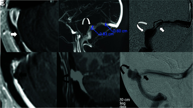
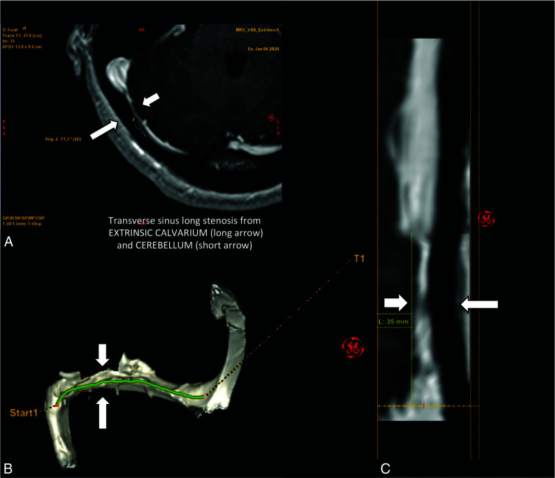
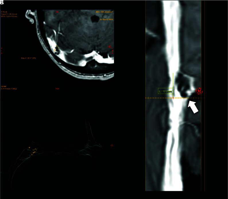
Background and purpose: Dural venous sinus stenosis has been associated with idiopathic intracranial hypertension and isolated venous pulsatile tinnitus. However, the utility of characterizing stenosis as intrinsic or extrinsic remains indeterminate. The aim of this retrospective study was to review preprocedural imaging of patients with symptomatic idiopathic intracranial hypertension and pulsatile tinnitus, classify the stenosis, and assess a trend between stenosis type and clinical presentation while reviewing the frequencies of other frequently seen imaging findings in these conditions.

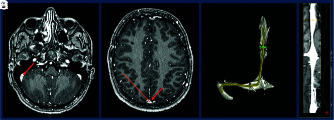
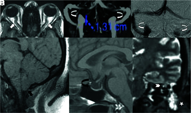
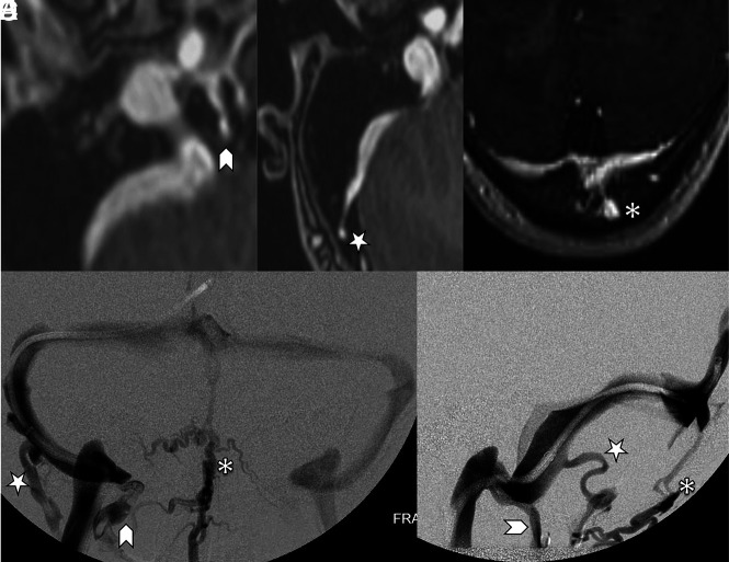
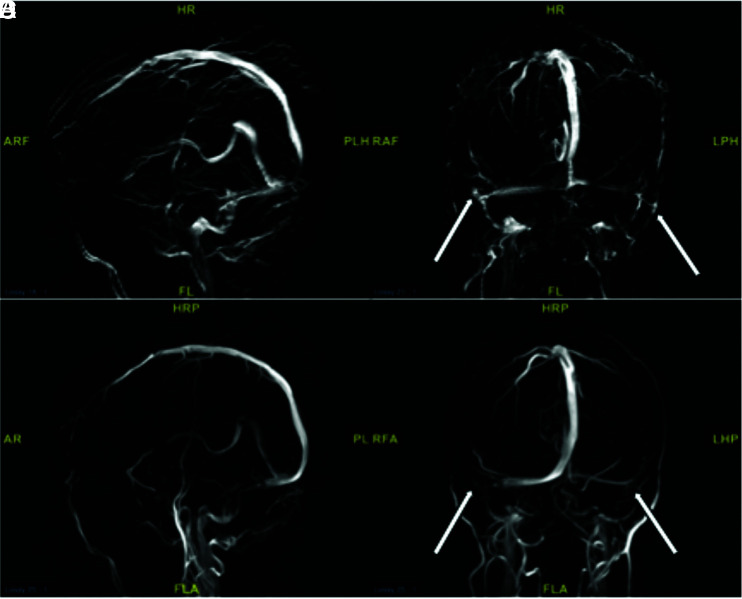
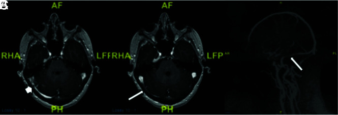
Materials and methods: MRVs of 115 patients with idiopathic intracranial hypertension and 43 patients with pulsatile tinnitus before venous sinus stent placement were reviewed. Parameters recorded included the following: intrinsic or extrinsic stenosis, prominent emissary veins, optic nerve tortuosity, cephalocele, sella appearance, poststenotic fusiform enlargement versus saccular venous aneurysm, and internal jugular bulb diverticula. χ2 cross-tabulation statistics were calculated and recorded for all data.

Results: Most patients with idiopathic intracranial hypertension (75 of 115 sinuses, 65%) had extrinsic stenosis, and most patients with pulsatile tinnitus (37 of 45 sinuses, 82%) had intrinsic stenosis. Marked optic nerve tortuosity was more common in idiopathic intracranial hypertension. Cephaloceles were rare in both cohorts, with an increased trend toward the presence in idiopathic intracranial hypertension. Empty sellas were more common in idiopathic intracranial hypertension. Cerebellar tonsils were similarly located at the foramen magnum level in both cohorts. Saccular venous aneurysms were more common in pulsatile tinnitus. Internal jugular bulb diverticula were similarly common in both cohorts.

Conclusions: In this cohort, most patients with idiopathic intracranial hypertension had extrinsic stenosis, and most patients with pulsatile tinnitus had intrinsic stenosis. Awareness and reporting of these subtypes may reduce the underrecognition of potential contributory stenoses in a given patient's idiopathic intracranial hypertension or pulsatile tinnitus.


Figures

FIG 1.
FIG 1.
A, Axial postcontrast MRV demonstrating extrinsic stenosis from the overlying cerebellum (short white arrow). B, Contrast-enhanced 3D-MRV image shows poststenotic sigmoid sinus enlargement (curved white arrow). C, Accompanying lateral venography confirms stenosis (white arrow) and sinus enlargement (curved white arrow) seen on the corresponding MRV. D, Separate axial postcontrast MRV shows intrinsic stenosis from arachnoid granulations (black arrow). E, A coronal postcontrast MRV sequence shows lateral sinus dehiscence with a venous aneurysm (curved black arrow). F, Accompanying frontal venography confirms stenosis (short black arrow) and a saccular aneurysm (curved black arrow) seen on the corresponding MRV.
FIG 2.
FIG 2.
A, 2-Click automatic vessel analysis start point selection in the sigmoid sinus (short arrow). B, 2-Click automatic vessel analysis end point selection (short arrow) in the superior sagittal sinus (long arrow). C, 3D volume-rendered vessel segmentation. D, Lumen view shows the straightened vessel segmentation.
FIG 3.
FIG 3.
Objective parameters implemented in recording corollary findings of both idiopathic intracranial hypertension and pulsatile tinnitus cohorts. A, Marked optic nerve tortuosity, with >50% of optic sheath width deviation noted relative to its expected straight path along the optic canal (white arrows). Bilateral  ≥ 5-mm internal jugular bulb diverticula, as seen on MRV (B) and catheter venography (C) images (curved white arrows). D, Cerebellar tonsil projecting 1–3 mm below the foramen magnum, referred to as ectopia (black arrow). E, Empty sella recorded if there is >75% loss of pituitary height (curved black arrow). F, Coronal T2 MR imaging demonstrates a left temporal lobe cephalocele through the tegmen tympani (arrowhead) and CSF in mastoid air cells (star).
FIG 4.
FIG 4.
Contrast-enhanced MRV images (A, B, and C) highlighting features of the emissary veins categorized in this study. A, A condylar vein is seen arising from the internal jugular vein bulb extending through the condylar canal (arrowhead). B, A mastoid emissary vein is seen arising from the sigmoid sinus traversing the mastoid foramen (star). C, An occipital emissary vein is seen arising from the torcula extending through the calvaria (asterisk). Conventional venography frontal (D) and lateral (E) images showcase the 3 emissary vein types categorized in this study. The condylar vein (arrowhead) extends inferiorly toward the vertebral plexus. The mastoid emissary vein (star) extends posteriorly and inferiorly to join the suboccipital plexus and external jugular vein. The occipital emissary vein (asterisk) drains inferiorly into the suboccipital plexus.
FIG 5.
FIG 5.
MR images showing extrinsic stenosis of the right transverse sinus. The short arrows point to right cerebellar parenchyma location, and the long arrows points to the occipital calvaria location. A, An axial contrast-enhanced MRV image. B, A 3D reconstruction image. C, A straight-vessel reformat of the right transverse-to-proximal sigmoid sinus from source contrast-enhanced axial images.
FIG 6.
FIG 6.
MR images showing intrinsic stenosis of the right transverse sinus. The short arrow points to a prominent arachnoid granulation situated inside the sinus. A, An axial contrast-enhanced MRV image, B, A 3D reconstruction image. C, A straight-vessel reformat of the right transverse-to-proximal sigmoid sinus from source contrast-enhanced axial images, noting orange shading of the arachnoid granulation and transparency of the remaining dural venous sinus.
FIG 7.
FIG 7.
Comparing TOF sagittal and coronal 3D reconstructions (A and B) and contrast-enhanced sagittal and coronal 3D reconstructions (C and D) from MRV in a patient with idiopathic intracranial hypertension. Note how the TOF images show the patient’s physiologic venous drainage due to properties of TOF imaging, showing only blood draining back to the patient’s heart (veins). Contrast-enhanced imaging, though crisper, shows arteries and veins in the same image. B and D, Arrows point to severe extrinsic stenoses in the bilateral transverse sinus–sigmoid sinus junctions, a common location for idiopathic intracranial hypertension stenosis. Such short-segment severe stenoses appear to simulate the abrupt narrowing commonly seen in short-segment intrinsic stenoses on these 3D reconstructions. However, review of source imaging would demonstrate brain parenchymal narrowing rather than primary arachnoid granulations producing the stenoses. HRP indicates head right posterior; PLH, posterior left head; RAF, right anterior foot; RA, right anterior; FL, foot left; PL, posterior left; RFA, right foot anterior; LHP, left head posterior; LPH, left posterior head; ARF, anterior right foot; AR, anterior right; FLA, foot left anterior; AF, anterior foot.
FIG 8.
FIG 8.
MR images demonstrating focal intrinsic stenosis just proximal to dominant extrinsic stenosis in this patient with idiopathic intracranial hypertension. The short arrow in A demonstrates focal arachnoid granulation. The long arrows in B and C demonstrate a primary extrinsic stenosis pattern. Despite the mixed presence of intrinsic and extrinsic stenoses, this was categorized as primary extrinsic stenosis. RHA indicates right head anterior; PH, posterior head; LFP, left foot posterior; AF, anterior foot; AR, anterior right; PL, posterior left; H, head; F, foot.

Comment in

References

    1. Dinkin MJ, Patsalides A. Venous sinus stenting for idiopathic intracranial hypertension: where are we now? Neurol Clin 2017;35:59–81 10.1016/j.ncl.2016.08.006 - DOI - PubMed
    1. Patsalides A, Oliveira C, Wilcox J, et al. . Venous sinus stenting lowers the intracranial pressure in patients with idiopathic intracranial hypertension. J Neurointerv Surg 2019;11:175–78 10.1136/neurintsurg-2018-014032 - DOI - PMC - PubMed
    1. Toscano S, Lo Fermo S, Reggio E, et al. . An update on idiopathic intracranial hypertension in adults: a look at pathophysiology, diagnostic approach and management. J Neurol 2020. May 27. [Epub ahead of print] 10.1007/s00415-020-09943-9 - DOI - PubMed
    1. Johnston I The historical development of the pseudotumor concept. Neurosurg Focus 2001;11:E2 10.3171/foc.2001.11.2.3 - DOI - PubMed
    1. Hewes D, Morales R, Raghavan P, et al. . Pattern and severity of transverse sinus stenosis in patients with pulsatile tinnitus associated with sigmoid sinus wall anomalies. Laryngoscope 2020;130:1028–33 10.1002/lary.28168 - DOI - PubMed