

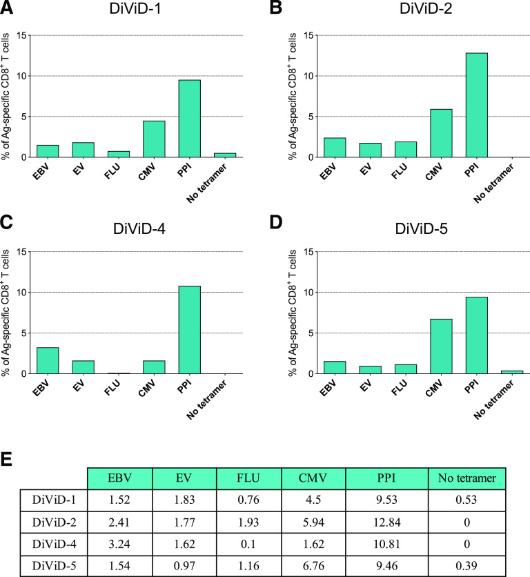
One in Ten CD8+ Cells in the Pancreas of Living Individuals With Recent-Onset Type 1 Diabetes Recognizes the Preproinsulin Epitope PPI15-24
- PMID: 33414250
- PMCID: PMC7897350
- DOI: 10.2337/db20-0908
One in Ten CD8+ Cells in the Pancreas of Living Individuals With Recent-Onset Type 1 Diabetes Recognizes the Preproinsulin Epitope PPI15-24
Abstract
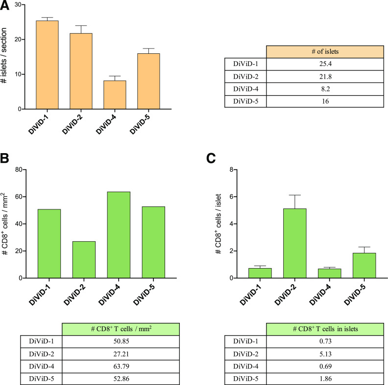
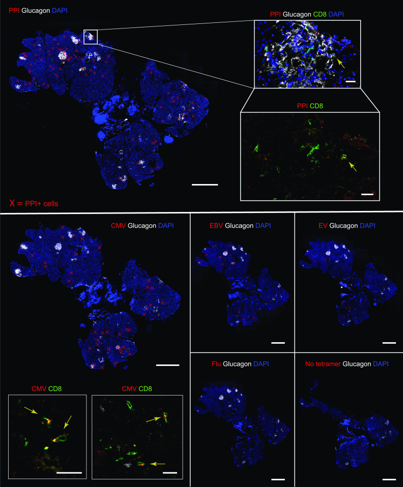
In type 1 diabetes (T1D), a lifelong autoimmune disease, T cells infiltrate the islets and the exocrine pancreas in high numbers. CD8+ T cells are the main cell type found in the insulitic lesion, and CD8+ T cells reactive against β-cell antigens have been detected in peripheral blood and in the pancreas of patients with short- or long-term disease. In the Diabetes Virus Detection (DiViD) study, researchers collected pancreatic tissue, by pancreatic tail resection, from living patients with recent-onset T1D. These tissues have been extensively studied by the scientific community, but the autoreactive nature of the T-cell infiltrate has remained unexplored. Our objective was to determine the number and localization of these cells in pancreas samples obtained through the DiViD study. Here, we demonstrate the presence of high frequencies of CD8+ T cells reactive against a highly relevant epitope derived from the preproinsulin signal peptide in pancreatic tissue samples from these donors. We also show the heterogeneity of islet distribution and CD8+ T-cell infiltration. Our findings contribute to the current limited existing knowledge of T-cell reactivity in the pancreas of donors with recent-onset T1D and indicate that antigen-specific therapies directed toward preproinsulin could have high clinical impact.
© 2021 by the American Diabetes Association.
Figures
Similar articles
-
Differential Insulitic Profiles Determine the Extent of β-Cell Destruction and the Age at Onset of Type 1 Diabetes.Diabetes. 2016 May;65(5):1362-9. doi: 10.2337/db15-1615. Epub 2016 Feb 8. Diabetes. 2016. PMID: 26858360
-
Human islet T cells are highly reactive to preproinsulin in type 1 diabetes.Proc Natl Acad Sci U S A. 2021 Oct 12;118(41):e2107208118. doi: 10.1073/pnas.2107208118. Proc Natl Acad Sci U S A. 2021. PMID: 34611019 Free PMC article.
-
Discovery of low-affinity preproinsulin epitopes and detection of autoreactive CD8 T-cells using combinatorial MHC multimers.J Autoimmun. 2011 Nov;37(3):151-9. doi: 10.1016/j.jaut.2011.05.012. Epub 2011 Jun 1. J Autoimmun. 2011. PMID: 21636247
-
Insulin and type 1 diabetes: immune connections.Eur J Endocrinol. 2013 Jan 17;168(2):R19-31. doi: 10.1530/EJE-12-0693. Print 2013 Feb. Eur J Endocrinol. 2013. PMID: 23065992 Review.
-
Narrowing in on the anti-β cell-specific T cells: looking 'where the action is'.Curr Opin Endocrinol Diabetes Obes. 2017 Apr;24(2):98-102. doi: 10.1097/MED.0000000000000323. Curr Opin Endocrinol Diabetes Obes. 2017. PMID: 28099204 Free PMC article. Review.
Cited by
-
A Humanized Mouse Strain That Develops Spontaneously Immune-Mediated Diabetes.Front Immunol. 2021 Oct 14;12:748679. doi: 10.3389/fimmu.2021.748679. eCollection 2021. Front Immunol. 2021. PMID: 34721418 Free PMC article.
-
The beta cell-immune cell interface in type 1 diabetes (T1D).Mol Metab. 2023 Dec;78:101809. doi: 10.1016/j.molmet.2023.101809. Epub 2023 Sep 20. Mol Metab. 2023. PMID: 37734713 Free PMC article. Review.
-
Monitoring immunomodulation strategies in type 1 diabetes.Front Immunol. 2023 Jun 6;14:1206874. doi: 10.3389/fimmu.2023.1206874. eCollection 2023. Front Immunol. 2023. PMID: 37346035 Free PMC article. Review.
-
Type 1 Diabetes: A Guide to Autoimmune Mechanisms for Clinicians.Diabetes Obes Metab. 2025 Aug;27 Suppl 6(Suppl 6):40-56. doi: 10.1111/dom.16460. Epub 2025 May 15. Diabetes Obes Metab. 2025. PMID: 40375390 Free PMC article. Review.
-
How benign autoimmunity becomes detrimental in type 1 diabetes.Proc Natl Acad Sci U S A. 2021 Nov 2;118(44):e2116508118. doi: 10.1073/pnas.2116508118. Proc Natl Acad Sci U S A. 2021. PMID: 34697240 Free PMC article. No abstract available.
References
-
- Krogvold L, Edwin B, Buanes T, et al. . Pancreatic biopsy by minimal tail resection in live adult patients at the onset of type 1 diabetes: experiences from the DiViD study. Diabetologia 2014;57:841–843 - PubMed
-
- Krogvold L, Wiberg A, Edwin B, et al. . Insulitis and characterisation of infiltrating T cells in surgical pancreatic tail resections from patients at onset of type 1 diabetes. Diabetologia 2016;59:492–501 - PubMed
-
- Gonzalez-Duque S, Azoury ME, Colli ML, et al. . Conventional and neo-antigenic peptides presented by β cells are targeted by circulating naïve CD8+ T cells in type 1 diabetic and healthy donors. Cell Metab 2018;28:946–960.e6 - PubMed
Publication types
MeSH terms
Substances
Associated data
Grants and funding
LinkOut - more resources
Full Text Sources
Other Literature Sources
Medical
Molecular Biology Databases
Research Materials
