Review
. 2021 Jan 8;6(1):8.
doi: 10.1038/s41392-020-00436-9.

Targeting TGFβ signal transduction for cancer therapy

Affiliations
Review

Targeting TGFβ signal transduction for cancer therapy

Sijia Liu et al. Signal Transduct Target Ther. .

Abstract

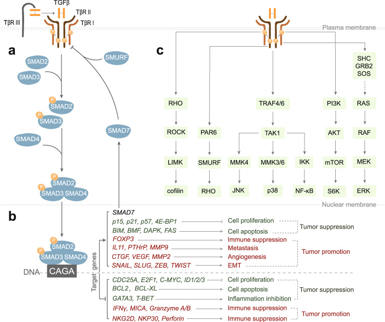
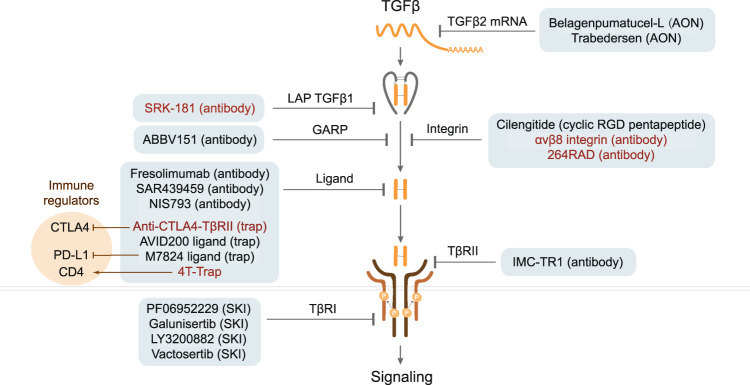
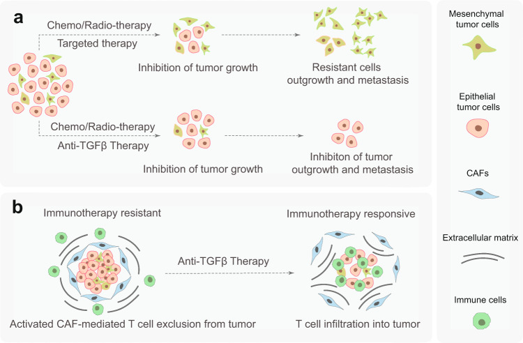
Transforming growth factor-β (TGFβ) family members are structurally and functionally related cytokines that have diverse effects on the regulation of cell fate during embryonic development and in the maintenance of adult tissue homeostasis. Dysregulation of TGFβ family signaling can lead to a plethora of developmental disorders and diseases, including cancer, immune dysfunction, and fibrosis. In this review, we focus on TGFβ, a well-characterized family member that has a dichotomous role in cancer progression, acting in early stages as a tumor suppressor and in late stages as a tumor promoter. The functions of TGFβ are not limited to the regulation of proliferation, differentiation, apoptosis, epithelial-mesenchymal transition, and metastasis of cancer cells. Recent reports have related TGFβ to effects on cells that are present in the tumor microenvironment through the stimulation of extracellular matrix deposition, promotion of angiogenesis, and suppression of the anti-tumor immune reaction. The pro-oncogenic roles of TGFβ have attracted considerable attention because their intervention provides a therapeutic approach for cancer patients. However, the critical function of TGFβ in maintaining tissue homeostasis makes targeting TGFβ a challenge. Here, we review the pleiotropic functions of TGFβ in cancer initiation and progression, summarize the recent clinical advancements regarding TGFβ signaling interventions for cancer treatment, and discuss the remaining challenges and opportunities related to targeting this pathway. We provide a perspective on synergistic therapies that combine anti-TGFβ therapy with cytotoxic chemotherapy, targeted therapy, radiotherapy, or immunotherapy.


Conflict of interest statement

The authors declare no competing interests.

Figures

Fig. 1
Fig. 1
Biphasic functions of TGFβ during tumor progression. TGFβ acts as a tumor suppressor in the initial stage of tumor progression by inducing cell cycle arrest and apoptosis of normal and pre-malignant epithelial cells. Upon activation of oncogenes and/or inactivation of tumor suppressor genes, tumor cells become insensitive to the TGFβ-induced cytostatic effects and undergo uncontrolled proliferation. TGFβ produced by tumor cells, fibroblasts, immune, and endothelial cells in the tumor microenvironment (TME) can trigger cancer cells to undergo an epithelial-to-mesenchymal transition (EMT). Thereby, late-stage cancer cells acquire the ability to escape from the primary niche, intravasate into the circulation, extravasate and localize to distant sites, and progress to form secondary tumors. Reciprocal TGFβ signaling between cancer cells and the TME contributes to cancer progression by activating cancer-associated fibroblasts (CAFs), stimulating angiogenesis, promoting protumor cytokine secretion, increasing extracellular matrix deposition, and evading an immune attack. In the metastatic sites, the mesenchymal tumor cells can undergo a mesenchymal-to-epithelial transition. Thus, thereby change back into an epithelial phenotype, which enables rapid outgrowth
Fig. 2
Fig. 2
A schematic representation of the activation of latent TGFβ. The pro-TGFβ precursor is synthesized in the rough endoplasmic reticulum. It consists of an N-terminal signal peptide, latency-associated peptide (LAP), and a mature C-terminal TGFβ fragment. After cleavage by the convertase furin in the Golgi complex, the LAP dimer binds to mature TGFβ noncovalently, preventing its binding to cell surface receptors, and forms the small latent complex (SLC). There are three major mechanisms for activation of latent TGFβ. a Proteases (e.g., cathepsin, plasmin, matrix metalloproteinase 9/14 (MMP9/14)) in the extracellular matrix (ECM) cleave LAP and release active TGFβ. Also, thrombospondin (TSP) can induce activation by direct binding to LAP. b SLC is anchored to the ECM proteins (e.g., fibronectin and fibrillin) via latent TGFβ-binding protein (LTBP) and forms the so-called large latent complex (LLC). Active TGFβ can be released by cell contraction upon the interaction between LAP and integrins. c SLC binds to glycoprotein A repetition predominant protein (GARP) on the cell surface and can also mediate the release of active TGFβ upon interaction with integrins
Fig. 3
Fig. 3
Schematic of the TGFβ-induced canonical SMAD and noncanonical signaling pathways. a TβRIII presents TGFβ to TβRII. Thereafter, ligand occupied TβRII recruits and phosphorylates TβRI to trigger intracellular TGFβ signaling pathways. In the canonical pathway, activated TβRI phosphorylates SMAD2/3 and stimulates the formation of heteromeric complexes with SMAD4. These complexes are translocated into the nucleus and regulate target gene expression. One of the TGFβ/SMAD-induced target genes is SMAD7, of which the gene product participates in a negative feedback loop to regulate the duration and intensity of TGFβ signaling by recruiting E3 ubiquitin ligase SMURF to TβRI. b TGFβ pathway target genes relevant for tumor suppression are listed in green, and the target genes that encode proteins involved in the tumor promotion are listed in red. c TGFβ can also activate many noncanonical pathways, including RHO, JNK, p38, NF-κB, AKT, and ERK signaling components
Fig. 4
Fig. 4
The functions of TGFβ upon the epithelial–mesenchymal transition (EMT). a Schematic of TGFβ mediation of the EMT process. b TGFβ promotes the EMT in non-transformed NAMRU mouse mammary gland (NMuMG) epithelial cells, as visualized by immunofluorescent staining of cells with anti-E-cadherin antibody (red) and phalloidin (green) to measure filamentous actin expression in the absence and after treatment with 5 ng/ml TGFβ3 for 48 h. The typical morphological change from epithelial- to fibroblast-like cells, decreased E-cadherin, and accumulated striated fibers are observed in the NMuMG cells upon TGFβ stimulation. c TGFβ promotes the migration of MDA-MB-231 human breast cancer cells, as determined via real-time imaging of a wound-healing scratch assay. Left, graph showing the time-lapse relative migration rate; right, images of the cells taken when the initial scratch was made (0 h) and after 24 and 48 h in the absence or presence of 5 ng/ml TGFβ3
Fig. 5
Fig. 5
Functions of TGFβ in the tumor microenvironment (TME). a TGFβ can activate/differentiate resident fibroblasts, mesenchymal stem cells, epithelial tumor cells, adipose tissue-derived stem cells, and endothelial cells into cancer-associated fibroblasts (CAFs) in the TME. b TGFβ promotes angiogenesis in the TME by acting directly and indirectly on endothelial cells stimulating their proliferation, migration, and sprouting. c TGFβ crucially suppresses the immune system by regulating the functions of immune cell populations in the TME. The specific actions of TGFβ are indicated in the boxes next to the different immune cells that are depicted
Fig. 6
Fig. 6
Schematic of strategies utilized in (pre)clinical trials targeting TGFβ signal transduction for cancer therapy. Various pharmacological interventions are grouped into the targeting of different TGFβ signaling components, that is, TGFβ mRNA, GARP/integrins that are involved in activation of latent TGFβ, and ligands that interact with TGFβ receptors and TβRI kinase activity. The promising new targeting molecules that have been studied in pre-clinical models are highlighted in red color. Different strategies for targeting TGFβ signaling, including antisense oligonucleotide (AON), neutralizing antibody (antibody), cyclic RGD pentapeptide, TGFβ ligand trap (trap), and small-molecule kinase inhibitor (SKI) are indicated. The immune regulatory targets (CTLA4/PD-L1/CD4) of the bispecific molecules that sequester TGFβ with a TβRII extracellular domain containing trap are highlighted in the orange circle
Fig. 7
Fig. 7
Sketch of synergistic combination therapies. a Chemo/radio/targeted therapy alone inhibits the growth of epithelial-like tumor cells, and in combination with anti-TGFβ therapy, invasive escape and resistance to these therapies are attenuated, and metastasis of mesenchymal tumor cells is restrained. b Activated CAFs mediated by high TGFβ activity suppresses immunotherapy efficacy by blocking T cell infiltration into tumors and inducing T cell dysfunction. In combination with anti-TGFβ therapy; however, T cell exclusion is inhibited, and the antitumor efficacy of the immunotherapy is improved

References

    1. David CJ, Massagué J. Contextual determinants of TGFβ action in development, immunity and cancer. Nat. Rev. Mol. Cell Biol. 2018;19:419–435. doi: 10.1038/s41580-018-0007-0. - DOI - PMC - PubMed
    1. Moustakas A, Heldin CH. The regulation of TGFβ signal transduction. Development. 2009;136:3699–3714. doi: 10.1242/dev.030338. - DOI - PubMed
    1. Batlle E, Massagué J. Transforming growth factor-β signaling in immunity and cancer. Immunity. 2019;50:924–940. doi: 10.1016/j.immuni.2019.03.024. - DOI - PMC - PubMed
    1. Derynck, R., Turley, S. J. & Akhurst, R. J. TGFβ biology in cancer progression and immunotherapy. Nat. Rev. Clin. Oncol. 10.1038/s41571-020-0403-1 (2020). - PMC - PubMed
    1. Flanders KC, et al. Quantitation of TGF-β proteins in mouse tissues shows reciprocal changes in TGF-β1 and TGF-β3 in normal vs neoplastic mammary epithelium. Oncotarget. 2016;7:38164–38179. doi: 10.18632/oncotarget.9416. - DOI - PMC - PubMed

Publication types

Substances