. 2021 Aug;35(8):2382-2398.
doi: 10.1038/s41375-020-01116-x. Epub 2021 Jan 7.

U2af1 is required for survival and function of hematopoietic stem/progenitor cells

Affiliations

U2af1 is required for survival and function of hematopoietic stem/progenitor cells

Avik Dutta et al. Leukemia. 2021 Aug.

Abstract

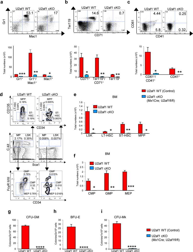
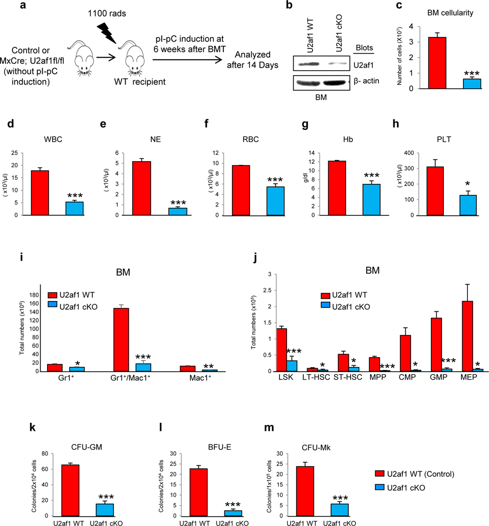
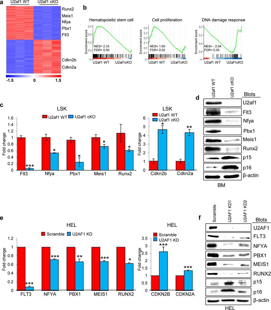
U2AF1 is involved in the recognition of the 3' splice site during pre-mRNA splicing. Mutations in U2AF1 are frequently observed in myelodysplastic syndromes. However, the role of wild-type U2AF1 in normal hematopoiesis has remained elusive. Using a novel conditional U2af1 knockout allele, we have found that deletion of U2af1 results in profound defects in hematopoiesis characterized by pancytopenia, ablation of hematopoietic stem/progenitor cells (HSPC) leading to bone marrow failure and early lethality in mice. U2af1 deletion impairs HSPC function and repopulation capacity. U2af1 deletion also causes increased DNA damage and reduced survival in hematopoietic progenitors. RNA sequencing analysis reveals significant alterations in the expression of genes related to HSC maintenance, cell proliferation, and DNA damage response-related pathways in U2af1-deficient HSPC. U2af1 deficiency also induces splicing alterations in genes important for HSPC function. This includes altered splicing and perturbed expression of Nfya and Pbx1 transcription factors in U2af1-deficient HSPC. Collectively, these results suggest an important role for U2af1 in the maintenance and function of HSPC in normal hematopoiesis. A better understanding of the normal function of U2AF1 in hematopoiesis is important for development of appropriate therapeutic approaches for U2AF1 mutant induced hematologic malignancies.


Conflict of interest statement

Conflict of interest

The authors declare that they have no conflict of interest.

Figures

Figure 1.
Figure 1.. Conditional deletion of U2af1 in mouse hematopoietic compartments results in fatal BM failure.
(a) Gene-targeting strategy for the U2af1 gene is depicted. The exon 2 of U2af1 is targeted in ES cells by inserting two LoxP sites between introns 1 and 2 of U2af1 gene to create U2af1 floxed allele. The U2af1 floxed allele can be deleted by the expression of Cre recombinase. (b) Immunoblot analysis for U2af1 protein showed efficient deletion of U2af1 in the BM of Mx1Cre;U2af1fl/fl mice (U2af1 cKO) after induction with pI-pC. Erk2 served as loading control. (c) qRT-PCR analysis also showed markedly reduced expression of U2af1 mRNA in U2af1 cKO mice BM (n=6) (**p<0.005). (d) Kaplan-Meier analysis showed significant decrease in survival of U2af1-deleted mice compared to control mice. Conditional U2af1-deleted mice died or became moribund within 10 −14 days after pI-pC (n=15 each group). Peripheral blood (e) white blood cell (WBC), (f) neutrophil (NE), (g) Red blood cell (RBC), (h) hemoglobin (Hb) and (i) platelet counts were assessed at 10 days after pI-pC induction in control and U2af1 cKO mice (n=10–12). (j) BM cells were ablated in U2af1 deleted mice compare to control mice 10 days after pI-pC (n=10 to 12). All data are shown as mean ± SEM. Student t-test was used to compare between two groups of mice (**p<0.005, ***p<0.0005). (k) Representative peripheral blood smears (1000X) from control and U2af1 cKO mice at 11 days after pI-pC injection are depicted. The U2af1 cKO blood smear shows severe anemia. H&E staining of the BM sections (40X and 500X) demonstrate pancytopenia and severe aplasia in U2af1 cKO mice BM.
Figure 2.
Figure 2.. Effects of U2af1 deletion on hematopoietic stem/progenitor cells.
Representative dot plots and bar graphs of flow cytometric analysis show significant decrease in (a) myeloid Gr1+/Mac1+), (b) erythroid (CD71+/Ter119+) and (c) megakaryocytic (CD61+/CD41+) populations in U2af1 cKO mice BM as compared to control mice 10 days after pI-pC (n=8 to 10) (*p<0.05, **p<0.005, ***p<0.0005). (d) Representative contour plots of flow cytometric analysis of LSK (Lin-Sca-1+c-kit+), LT-HSC (Lin-Sca-1+c-kit+CD34-CD135-), ST-HSC (Lin-Sca-1+c-kit+CD34+CD135-), MPP (Lin-Sca-1+c-kit+CD34+CD135+), CMP (Lin-Sca-1-c-kit+CD34+FcγRII/IIlow), GMP (Lin-Sca-1-c-kit+CD34+FcγRII/IIhigh) and MEP (Lin-Sca-1-c-kit+CD34-FcγRII/III-) in the BM of control and U2af1 cKO mice BM at 10 days after pI-pC. (e) Total numbers of LSK, LT-HSC, ST-HSC and MPP in the BM of control and U2af1 cKO mice are shown in bar graphs as mean ± SEM (n=8 to 10). (f) Total numbers of CMP, GMP and MEP in the BM of control and U2af1 cKO mice are shown in bar graphs as mean ± SEM (n=8 to 10). (*p<0.05, **p<0.005, ***p<0.0005). (g-h) Hematopoietic progenitor colony assays. Total BM cells (2 × 10⁴) from control WT and U2af1 cKO mice (n=6) were plated in methylcellulose medium (MethoCult 3434) with cytokines. CFU-GM and BFU-E colonies were scored 7 days after plating. (i) BM cells (1 × 10⁵) from control and U2af1 cKO mice (n=6) were plated into collagen-based MegaCult medium supplemented with IL-3, IL-6, IL-11, and Tpo. Megakaryocytic (CFU-Mk) colonies were assessed after culturing for 8 days. All data are shown as mean ± SEM (****p<0.00005 by student t-test).
Figure 3.
Figure 3.. BM failure observed in U2af1 deficient mice is cell autonomous.
(a) Experimental design for cell autonomous bone marrow transplantation (BMT) assay. BM cells (1×10⁶) were harvested from uninduced Mx1Cre;U2af1fl/fl and control mice, and transplanted into lethally irradiated wild type C57BL/6J recipient mice. Deletion of U2af1 was induced in donor-derived hematopoietic cells by injecting 3 doses of pI-pC at 6 weeks after BMT. Mice were analyzed 14 days after first pI-pC injection. (b) Immunoblot analysis showed efficient deletion of U2af1 in the BM of Mx1Cre;U2af1fl/fl recipient animals after induction with pI-pC. β-actin was used as a loading control. (c) BM cellularity was markedly reduced in U2af1-deficient mice compared to control mice 14 days after pI-pC (n=7 to 10). Peripheral blood (d) White blood cell (WBC), (e) neutrophil (NE), (f) Red blood cell (RBC), (g) hemoglobin (Hb) and (h) platelet (PLT) counts were assessed at 11 days after pI-pC induction in control and U2af1-deficient BMT mice (n=10 to 20). (i) Bar graphs of flow cytometric analysis show significant decrease in myeloid Gr1+/Mac1+) precursors in the BM of U2af1-deficient BMT mice as compared to control recipient mice at 14 days after pI-pC induction. (j) Bar graphs showing the total numbers of LSK, LT-HSC, ST-HSC, MPP, CMP, GMP and MEP in the BM of control and U2af1-deficient BMT mice. The data are presented as mean ± SEM (control, n=7; U2af1 cKO=10) (*p<0.05, **p<0.005, ***p<0.0005). (k-l) BM cells (2 × 10⁴) from control and U2af1-deficient BMT mice (n=5) were plated in methylcellulose medium supplemented with cytokines. CFU-GM and BFU-E colonies were scored 7 days after plating. (m) BM cells (1 × 10⁵) from control and U2af1-deficient BMT mice (n=5) were plated into collagen-based MegaCult medium supplemented with IL-3, IL-6, IL-11, and Tpo. CFU-Mk colonies were assessed after culturing for 8 days. All data are shown as mean ± SEM (***p<0.0005).
Figure 4.
Figure 4.. Defective stem cell function in U2af1-deficient mice.
(a) Competitive reconstitution assay. BM cells (5×10⁵) from uninduced CD45.2+ Mx1Cre;U2af1fl/fl (U2af1 cKO) or littermate control mice were mixed with CD45.1+ wild type mice BM (5×10⁵) at a 1:1 ratio and transplanted into lethally irradiated CD45.1+ recipient mice. Four weeks after BMT, recipient mice were treated with 3 doses of pI-pC to induce U2af1 deletion after hematopoietic reconstitution. The recipient mice were analyzed at 16 weeks after pI-pC injection. (b) The percentages of donor-derived CD45.2+ cells, (c) percentages of CD45.2+ Gr-1+ myeloid cells (d) percentages of CD45.2+ B220+ B cells and (e) percentages of CD45.2+ Tcrβ+ T cells in the peripheral blood of recipient animals at 2 and 16 weeks after pI-pC induction are shown in bar graphs. (f) Representative contour plots of flow cytometric analysis and the percentages of donor-derived CD45.2+ total BM cells, (g) percentages of CD45.2+ Gr-1+ myeloid cells and (h) percentages of CD45.2+ LSK cells in the BM of recipient animals at 16 weeks after pI-pC induction are shown; bar graphs represent mean ± SEM. (Control, n=10; U2af1 cKO=10). Student t-test was used to compare between two groups of mice (**p<0.005, ***p<0.0005 and ****p<0.00005).
Figure 5.
Figure 5.. Deletion of U2af1 leads to DNA damage and apoptosis in hematopoietic cells.
(a) Annexin V/DAPI staining was performed on hematopoietic progenitors and precursor cells from U2af1 cKO and control mice BM at 8 days after pI-pC induction and apoptosis was measured by flow cytometry. Bar graphs showing increased percentage of apoptotic cells in the total BM, c-Kit+ progenitors, Gr1+ myeloid, CD71+ erythroid and CD41+ megakaryocyte cells in U2af1 cKO mice compared to control mice. Data are presented as mean ± SEM (control n=6; U2af1 cKO n=6) (***p<0.0005, ****p<0.00005). (b) Representative images from the Image stream flow imaging show increased γ-H2AX+ cells in the BM of U2af1 cKO mice compared with WT control mice. γ-H2AX (red) and DAPI (blue). (c) Image stream flow imaging show marked shift in the fluorescence intensity of γ-H2AX+ cells suggesting increased DNA damage in U2af1 cKO BM. (d) Bar graphs showing quantification of γ-H2AX+ cells in the total BM, Gr1+ myeloid and CD71+ erythroid cells in control and U2af1 cKO mice BM at 8 days after pI-pC administration. Data are presented as mean ± SEM (control n=6; U2af1 cKO n=6) (*p<0.05, **p<0.005, ****p<0.00005). (e) Representative immunofluorescence images of BM sections showing increased γ-H2AX (serine 139) staining in U2af1 cKO mice BM as compared to control mice BM. γ-H2AX (green) and DAPI (blue) Scale bars, 10μm. (f) Immunoblot analyses showing increased phosphorylation of H2AX at serine 139 (γ-H2AX) and CHK1 at serine 345, and increased ubiquitination of H2A at lysine 119 (H2AK119Ub) in U2af1 cKO mice BM cells compared with WT control BM cells. β-Actin was used as a loading control. (g) Immunoblot analyses showing increased phosphorylation of H2AX at serine 139 (γ-H2AX) and CHK1 at serine 345, and increased ubiquitination of H2AK119 (H2AK119Ub) in HEL cells upon U2AF1 knockdown. β-Actin was used as a loading control. (h) Representative immunofluorescence images of BM sections showing increased R-loop signals by S9.6 staining in U2af1 cKO mice BM as compared to control mice BM. S9.6 (green) and DAPI (blue) Scale bars, 15μm. (i) R-loop signals detected by immunofluorescence staining with S9.6 in BM Lin- cells from control and U2af1 cKO mice at 7 days after pI-pC induction. S9.6 (green) and DAPI (blue) Scale bars, 15μm. (j) Quantification of S9.6+ cells from control and U2af1 cKO mice BM Lin- cells is presented in bar graphs. Data are presented as mean ± SEM (control n=3; U2af1 cKO n=3; more than 100 cells were analyzed) (***p<0.0005).
Figure 6.
Figure 6.. Effect of U2af1 deletion on gene expression profile in HSPC.
(a) Heat map showing significantly up-regulated and down-regulated (p-adj <0.05; fold change >1.5 fold and <−1.5 fold) genes in U2af1-deficient LSK cells compared with control LSK cells in BMT mice. (b) Gene-set enrichment analyses show significant alterations of genes related to hematopoietic stem cells, cell proliferation and DNA damage response pathways in U2af1-deficient LSK cells compared with control LSK cells. (c) Relative expression of Flt3, Nfya, Pbx1, Meis1, Runx2, Cdkn2a (p16), and Cdkn2b (p15) mRNA was determined in control WT and U2af1-deficient LSK cells by RT-qPCR and normalized with Hprt expression. (d) Immunoblot analyses show decreased protein levels of Flt3, Nfya, Pbx1, Meis1 and Runx2 and increased levels of p15 and p16 in U2af1 cKO BM compared with control BM. (e) Validation of U2AF1 targets using lentiviral U2AF1 knockdown in HEL cells. Relative expression of FLT3, NFYA, PBX1, MEIS1, RUNX2, CDKN2A (p16), and CDKN2B (p15) was assessed by RT-qPCR and normalized by HPRT expression. Data are shown in bar graphs as mean ± SEM (n =4; *p<0.05, **p<0.005, ***p<0.0005). (f) Immunoblot analyses show decreased protein levels of FLT3, NFYA, PBX1, MEIS1 and RUNX2 and increased levels of p15 and p16 in U2AF1 knockdown HEL cells. β-Actin was used as a loading control.
Figure 7.
Figure 7.. Effect of U2af1 deletion on RNA splicing.
(a) Number and type of alternative splicing events in U2af1-deficient LSK cells compared with control LSK cells. (b) Inclusion levels of cassette exons (exon skipping/inclusion) in U2af1-deficient LSK cells compared with control LSK cells are depicted. Red and blue colored dots represent individual cassette exons that are significantly more included (red) or excluded/skipped (blue) in U2af1-deficient versus control LSK cells (>5% inclusion level differences and FDR <0.05). (c) Venn diagrams comparing gene expression changes and splicing events in U2af1-deficient LSK cells compared with control LSK cells. (d-e) Sashimi plots, RT-PCR and gel electrophoresis analyses confirmed altered splicing events in Nfya and Pbx1. % of long isoform was significantly decreased in U2af1 cKO LSK cells compared with control LSK cells for both Nfya and Pbx1(*p<0.05, **p<0.005). (f-g) Ectopic expression of Nfya or Pbx1 by lentiviral transduction partially rescues the defects in hematopoietic progenitor colony formation in U2af1-deficient BM progenitors. Transduced cells were selected using puromycin and plated in methylcellulose medium containing complete cytokines or Epo (3U/ml). Myeloid progenitor colonies (CFU-GM and CFU-GEMM) and erythroid (CFU-E) colonies were counted. Results are expressed as percentage of controls (n=4; **p<0.005, ***p<0.0005).

References

    1. Yoshida K, Sanada M, Shiraishi Y, Nowak D, Nagata Y, Yamamoto R, et al. Frequent Pathway Mutations of Splicing Machinery in Myelodysplasia. Nature, 2011. 478(7367):64–69. - PubMed
    1. Papaemmanuil E, Cazzola M, Boultwood J, Malcovati L, Vyas P, Bowen D, et al. Somatic SF3B1 Mutation in Myelodysplasia With Ring Sideroblasts. N Engl J Med, 2011. 365(15): 1384–95. - PMC - PubMed
    1. Graubert TA, Shen D, Ding L, Okeyo-Owuor T, Lunn CL, Shao J, et al. Recurrent mutations in the U2AF1 splicing factor in myelodysplastic syndromes. Nat Genet., 2012. 44(1): 53–57. - PMC - PubMed
    1. Thol F, Kade S, Schlarmann C, Löffeld P, Morgan M, Krauter J, et al. Frequency and Prognostic Impact of Mutations in SRSF2, U2AF1, and ZRSR2 in Patients With Myelodysplastic Syndromes. Blood, 2012. 119(15): 3578–84. - PubMed
    1. Damm F, Kosmider O, Gelsi-Boyer V, Renneville A, Carbuccia N, Hidalgo-Curtis C, et al. Groupe Francophone des Myélodysplasies., Mutations Affecting mRNA Splicing Define Distinct Clinical Phenotypes and Correlate With Patient Outcome in Myelodysplastic Syndromes. Blood, 2012. 119(14): 3211–18. - PubMed

Publication types