

The Osteogenetic Potential of Chitosan Coated Implant: An In Vitro Study
- PMID: 33414580
- PMCID: PMC7772811
- DOI: 10.46582/jsrm.1602008
The Osteogenetic Potential of Chitosan Coated Implant: An In Vitro Study
Abstract
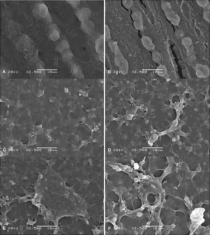
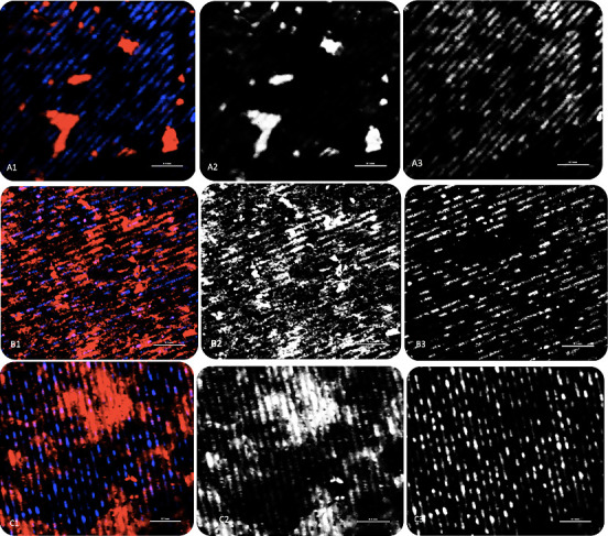
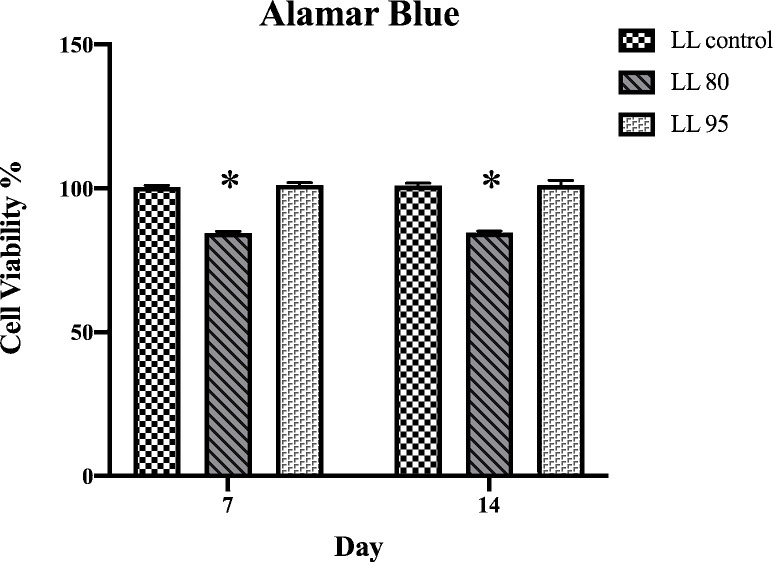
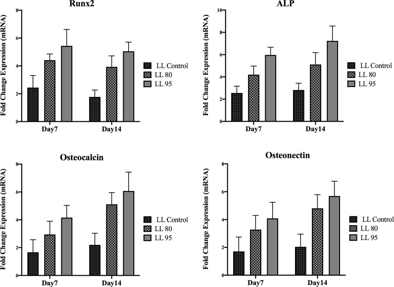
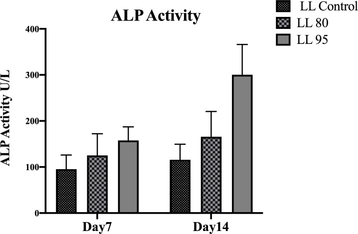
Objective: Chitosan is a promising polymer that has been used for coating dental implants. However, research concerning coatings with implant surfaces other than commercially pure titanium is limited. Therefore, this study aims to clarify the chitosan material's effect with two degrees of deacetylation (DDA) as coatings for laser surface microtopographic implants. Methods: Sixty-three Laser-Lok (LL) implant discs were divided into three groups (21 in each group), and two groups were coated with either 80 or 95 DDA chitosan. The groups were categorized as LL 95, LL 80, or LL control. Then, hMSC-TERT 20 cells were used to evaluate the cell morphology, viability, and osteogenic capacity of the chitosan material 7 and 14 days after culture. Two-way ANOVA followed by one-way analysis of variance (ANOVA) and Tukey's post hoc test were used. Results: All samples were biocompatible and allowed cell attachment. However, cell spreading and attachment were noticeably increased in the LL 95 group. There was a significant increase in the expression of osteogenic markers in chitosan-coated samples compared to the control group. The 95 DDA-coated group exhibited higher ALP, Runx2, osteocalcin, and osteonectin expression compared to the 80 DDA and control groups on days 7 and 14. Conclusion: A high DDA of chitosan promotes biomineralization and osteoblast formation. Therefore, this combination of laser surface and chitosan can enhance future dental implant healing processes and osseointegration.
Keywords: Chitosan; Implant coating; Laser lock.
Copyright © Journal of Stem Cells and Regenerative Medicine.
Conflict of interest statement
None
Figures

Similar articles
-
Role of chitosan in titanium coatings. trends and new generations of coatings.Front Bioeng Biotechnol. 2022 Jul 22;10:907589. doi: 10.3389/fbioe.2022.907589. eCollection 2022. Front Bioeng Biotechnol. 2022. PMID: 35935477 Free PMC article. Review.
-
Mechanical property, degradation rate, and bone cell growth of chitosan coated titanium influenced by degree of deacetylation of chitosan.J Biomed Mater Res B Appl Biomater. 2008 Jul;86(1):245-52. doi: 10.1002/jbm.b.31012. J Biomed Mater Res B Appl Biomater. 2008. PMID: 18161778
-
Contact angle, protein adsorption and osteoblast precursor cell attachment to chitosan coatings bonded to titanium.J Biomater Sci Polym Ed. 2003;14(12):1401-9. doi: 10.1163/156856203322599734. J Biomater Sci Polym Ed. 2003. PMID: 14870943
-
Effects of magnesium-substituted nanohydroxyapatite coating on implant osseointegration.Clin Oral Implants Res. 2013 Aug;24 Suppl A100:34-41. doi: 10.1111/j.1600-0501.2011.02362.x. Epub 2011 Dec 6. Clin Oral Implants Res. 2013. PMID: 22145854
-
Significance of osteogenic surface coatings on implants to enhance osseointegration under osteoporotic-like conditions.Implant Dent. 2014 Dec;23(6):679-86. doi: 10.1097/ID.0000000000000161. Implant Dent. 2014. PMID: 25290281 Review.
Cited by
-
Cell Adhesion and Initial Bone Matrix Deposition on Titanium-Based Implants with Chitosan-Collagen Coatings: An In Vitro Study.Int J Mol Sci. 2023 Mar 2;24(5):4810. doi: 10.3390/ijms24054810. Int J Mol Sci. 2023. PMID: 36902249 Free PMC article.
-
Chitosan nanoparticle applications in dentistry: a sustainable biopolymer.Front Chem. 2024 Apr 10;12:1362482. doi: 10.3389/fchem.2024.1362482. eCollection 2024. Front Chem. 2024. PMID: 38660569 Free PMC article. Review.
-
Recent Advancements in Chitosan-Based Biomaterials for Wound Healing.J Funct Biomater. 2025 Jan 30;16(2):45. doi: 10.3390/jfb16020045. J Funct Biomater. 2025. PMID: 39997579 Free PMC article. Review.
-
Bacterial Cellulose-A Remarkable Polymer as a Source for Biomaterials Tailoring.Materials (Basel). 2022 Jan 29;15(3):1054. doi: 10.3390/ma15031054. Materials (Basel). 2022. PMID: 35160997 Free PMC article. Review.
-
Role of chitosan in titanium coatings. trends and new generations of coatings.Front Bioeng Biotechnol. 2022 Jul 22;10:907589. doi: 10.3389/fbioe.2022.907589. eCollection 2022. Front Bioeng Biotechnol. 2022. PMID: 35935477 Free PMC article. Review.
References
-
- Albrektsson T, Brånemark PI, Hansson HA, Lindström J. Osseointegrated titanium implants. Requirements for ensuring a long-lasting, direct bone-to-implant anchorage in man. Acta Orthop Scand. 1981;52((2)):155–70. - PubMed
-
- Chrcanovic BR, Kisch J, Albrektsson T, Wennerberg A. Factors Influencing Early Dental Implant Failures. J Dent Res. 2016;95((9)):995–1002. - PubMed
-
- Soskolne WA, Cohen S, Sennerby L, Wennerberg A, Shapira L. The effect of titanium surface roughness on the adhesion of monocytes and their secretion of TNF-alpha and PGE2. Clin Oral Implants Res. 2002;13((1)):86–93. - PubMed
-
- Junker R, Dimakis A, Thoneick M, Jansen JA. Effects of implant surface coatings and composition on bone integration: a systematic review. Clin Oral Implants Res. 2009;20(Suppl 4):185–206. - PubMed
-
- Deppe H, Wolff C, Bauer F, Ruthenberg R, Sculean A, Mücke T. Dental implant surfaces after insertion in bone: an in vitro study in four commercial implant systems. Clin Oral Investig. 2018;22((3)):1593–1600. - PubMed
LinkOut - more resources
Full Text Sources