Adiponectin Interacts In-Vitro With Cementoblasts Influencing Cell Migration, Proliferation and Cementogenesis Partly Through the MAPK Signaling Pathway
- PMID: 33414717
- PMCID: PMC7783624
- DOI: 10.3389/fphar.2020.585346
Adiponectin Interacts In-Vitro With Cementoblasts Influencing Cell Migration, Proliferation and Cementogenesis Partly Through the MAPK Signaling Pathway
Abstract
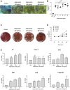
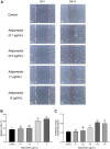
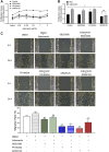
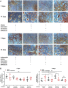
Current clinical evidences suggest that circulating Adipokines such as Adiponectin can influence the ratio of orthodontic tooth movement. We aimed to investigate the effect that Adiponectin has on cementoblasts (OCCM-30) and on the intracellular signaling molecules of Mitogen-activated protein kinase (MAPK). We demonstrated that OCCM-30 cells express AdipoR1 and AdipoR2. Alizarin Red S staining revealed that Adiponectin increases mineralized nodule formation and quantitative AP activity in a dose-dependent manner. Adiponectin up-regulates the mRNA levels of AP, BSP, OCN, OPG, Runx-2 as well as F-Spondin. Adiponectin also increases the migration and proliferation of OCCM-30 cells. Moreover, Adiponectin induces a transient activation of JNK, P38, ERK1/2 and promotes the phosphorylation of STAT1 and STAT3. The activation of Adiponectin-mediated migration and proliferation was attenuated after pharmacological inhibition of P38, ERK1/2 and JNK in different degrees, whereas mineralization was facilitated by MAPK inhibition in varying degrees. Based on our results, Adiponectin favorably affect OCCM-30 cell migration, proliferation as well as cementogenesis. One of the underlying mechanisms is the activation of MAPK signaling pathway.
Keywords: MAPK; adiponectin; cementoblasts; cementogenesis; migration; proliferation.
Copyright © 2020 Yong, von Bremen, Ruiz-Heiland and Ruf.
Conflict of interest statement
The authors declare that the research was conducted in the absence of any commercial or financial relationships that could be construed as a potential conflict of interest.
Figures


Similar articles
-
Ciliary Neurotrophic Factor (CNTF) Inhibits In Vitro Cementoblast Mineralization and Induces Autophagy, in Part by STAT3/ERK Commitment.Int J Mol Sci. 2022 Aug 18;23(16):9311. doi: 10.3390/ijms23169311. Int J Mol Sci. 2022. PMID: 36012576 Free PMC article.
-
Adiponectin as Well as Compressive Forces Regulate in vitro β-Catenin Expression on Cementoblasts via Mitogen-Activated Protein Kinase Signaling Activation.Front Cell Dev Biol. 2021 Apr 28;9:645005. doi: 10.3389/fcell.2021.645005. eCollection 2021. Front Cell Dev Biol. 2021. PMID: 33996803 Free PMC article.
-
Effect of Low-Level Er: YAG (2940 nm) laser irradiation on the photobiomodulation of mitogen-activated protein kinase cellular signaling pathway of rodent cementoblasts.Front Biosci (Landmark Ed). 2022 Feb 14;27(2):62. doi: 10.31083/j.fbl2702062. Front Biosci (Landmark Ed). 2022. PMID: 35227005
-
Intermittent parathyroid hormone (PTH) promotes cementogenesis and alleviates the catabolic effects of mechanical strain in cementoblasts.BMC Cell Biol. 2017 Apr 20;18(1):19. doi: 10.1186/s12860-017-0133-0. BMC Cell Biol. 2017. PMID: 28427342 Free PMC article.
-
Effect of interleukin-33 on cementoblast-mediated cementum repair during orthodontic tooth movement.Arch Oral Biol. 2020 Apr;112:104663. doi: 10.1016/j.archoralbio.2020.104663. Epub 2020 Jan 13. Arch Oral Biol. 2020. PMID: 31986333
Cited by
-
PD-L1, a Potential Immunomodulator Linking Immunology and Orthodontically Induced Inflammatory Root Resorption (OIIRR): Friend or Foe?Int J Mol Sci. 2022 Sep 27;23(19):11405. doi: 10.3390/ijms231911405. Int J Mol Sci. 2022. PMID: 36232704 Free PMC article. Review.
-
Ciliary Neurotrophic Factor (CNTF) and Its Receptors Signal Regulate Cementoblasts Apoptosis through a Mechanism of ERK1/2 and Caspases Signaling.Int J Mol Sci. 2022 Jul 28;23(15):8335. doi: 10.3390/ijms23158335. Int J Mol Sci. 2022. PMID: 35955469 Free PMC article.
-
Selection and validation of reference gene for RT-qPCR studies in co-culture system of mouse cementoblasts and periodontal ligament cells.BMC Res Notes. 2022 Feb 15;15(1):57. doi: 10.1186/s13104-022-05948-x. BMC Res Notes. 2022. PMID: 35168676 Free PMC article.
-
Immunorthodontics: PD-L1, a Novel Immunomodulator in Cementoblasts, Is Regulated by HIF-1α under Hypoxia.Cells. 2022 Jul 30;11(15):2350. doi: 10.3390/cells11152350. Cells. 2022. PMID: 35954195 Free PMC article.
-
Transcriptome Profile of Membrane and Extracellular Matrix Components in Ligament-Fibroblastic Progenitors and Cementoblasts Differentiated from Human Periodontal Ligament Cells.Genes (Basel). 2022 Apr 8;13(4):659. doi: 10.3390/genes13040659. Genes (Basel). 2022. PMID: 35456465 Free PMC article.
References
-
- Benedix F., Westphal S., Patschke R., Granowski D., Luley C., Lippert H., et al. (2011). Weight loss and changes in salivary ghrelin and adiponectin: comparison between sleeve gastrectomy and Roux-en-Y gastric bypass and gastric banding. Obes. Surg 21 (5), 616–624. 10.1007/s11695-011-0374-5 - DOI - PubMed
LinkOut - more resources
Full Text Sources
Research Materials
Miscellaneous

