Review
. 2020 Dec 22:11:617459.
doi: 10.3389/fphys.2020.617459. eCollection 2020.

Autonomic Modulation for Cardiovascular Disease

Affiliations
Review

Autonomic Modulation for Cardiovascular Disease

Joseph Hadaya et al. Front Physiol. .

Abstract

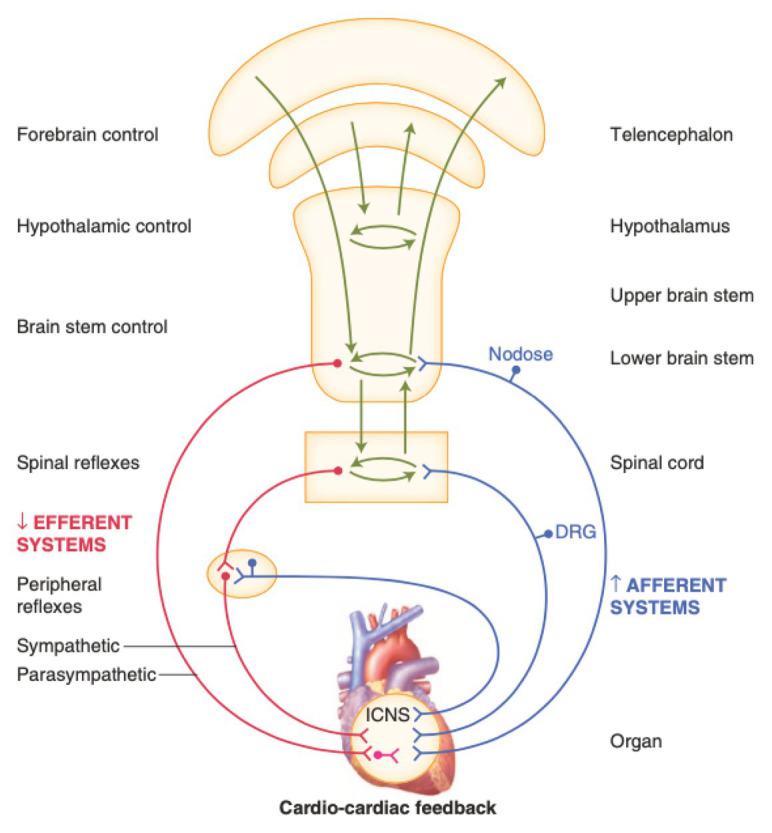
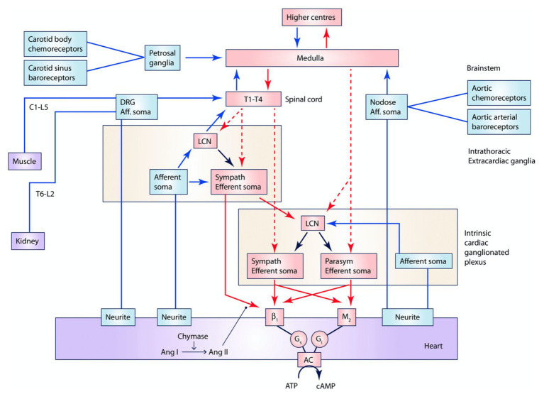
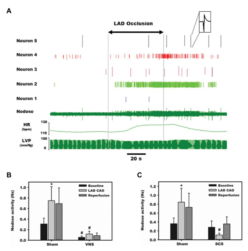
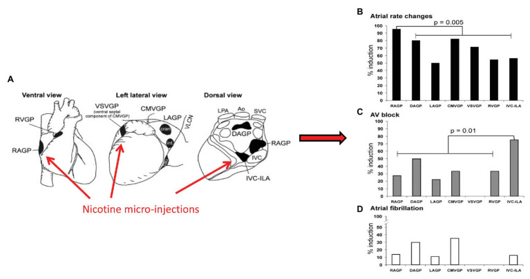
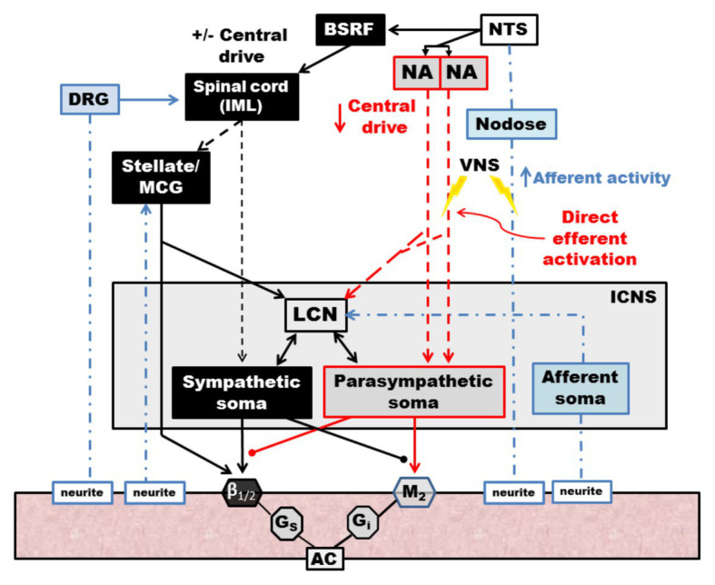
Dysfunction of the autonomic nervous system has been implicated in the pathogenesis of cardiovascular disease, including congestive heart failure and cardiac arrhythmias. Despite advances in the medical and surgical management of these entities, progression of disease persists as does the risk for sudden cardiac death. With improved knowledge of the dynamic relationships between the nervous system and heart, neuromodulatory techniques such as cardiac sympathetic denervation and vagal nerve stimulation (VNS) have emerged as possible therapeutic approaches for the management of these disorders. In this review, we present the structure and function of the cardiac nervous system and the remodeling that occurs in disease states, emphasizing the concept of increased sympathoexcitation and reduced parasympathetic tone. We review preclinical evidence for vagal nerve stimulation, and early results of clinical trials in the setting of congestive heart failure. Vagal nerve stimulation, and other neuromodulatory techniques, may improve the management of cardiovascular disorders, and warrant further study.

Keywords: arrhythmia; autonomic nervous system; heart failure; myocardial infaraction; neurocardiology; neuromodulation; sympathectomy; vagus nerve.


Conflict of interest statement

University of California, Los Angeles has patents developed by JLA relating to cardiac neural diagnostics and therapeutics. JLA is a co-founder of NeuCures, Inc. The remaining author declares that the research was conducted in the absence of any commercial or financial relationships that could be construed as a potential conflict of interest.

Figures

Figure 1
Figure 1
Neural regulation of cardiac function involves multiple nested feedback loops at the level of the heart, peripheral ganglia, and central nervous system. Afferent systems (blue) are mediated through the intrinsic cardiac nervous system (ICNS), dorsal root ganglia (DRG), and nodose ganglia. Efferent systems (red) involve sympathetic, parasympathetic, and local circuit neuron (LCNs). DRG, dorsal root ganglia. ICNS, intrinsic cardiac nervous system. Adapted from Jänig (2014) with permission.
Figure 2
Figure 2
Interactions between central nervous system, peripheral ganglia, and the ICNS. Multiple interdependent feedback loops regulate regional cardiac mechanical and electrical function. At the level of the heart, sensory (blue) neurons provide input directly and indirectly through the ICNS, peripheral ganglia, and spinal cord, which are capable of engaging cardiocentric reflexes. Converging inputs result in activation of adrenergic or muscarinic receptors as well as receptors for peptide co-transmitters such as neuropeptide Y (not displayed). Preganglionic sympathetic and parasympathetic efferent fibers project (red, dashed) to the ICNS, including to LCNs, and postganglionic sympathetic fibers (red, solid) also directly to the myocardium. The ICNS acts as the final common pathway for cardiac control, though interactions occur at all levels. Input from chemoreceptors, baroreceptors, as well as neurohormonal factors such as angiotensin II and circulating epinephrine (not displayed), also modulate cardiac function. Ang I, angiotensin I. Ang II, angiotensin II. LCN, local circuit neuron. Adapted from Ardell et al. (2016) with permission.
Figure 3
Figure 3
Afferent transduction of ischemia is modulated directly by parasympathetic input as well as remotely. Representative recordings from the nodose ganglia, one of the major parasympathetic afferent ganglia, during occlusion of the left anterior descending (LAD) coronary artery. (A) Activity from five neurons displays substantial heterogeneity in baseline firing frequency and responses to ischemia and reperfusion. (B) Normalized, summative nodose activity at baseline, with LAD artery occlusion, and reperfusion, with and without vagal nerve stimulation. In all settings, animals receiving vagal nerve stimulation (VNS) displayed marked reductions in nodose activity. (C) Spinal cord stimulation also reduces nodose neural activity in response to ischemia stress. Adapted from Salavatian et al. (2017a) with permission. *p < 0.05 from baseline; #p < 0.05 from sham.
Figure 4
Figure 4
Aggregates of afferent, efferent, and interneurons located within epicardial fat pads comprise the ICNS. Although each ganglionated plexus has a preferred sphere of influence, substantial overlap exists. (A) Ventral, left lateral and dorsal views of the heart demonstrating common atrial and ventricular ganglionated plexi. Selected electrophysiologic effects of nicotine microinjections into major atrial and ventricular ganglionated plexi, (B) Heart rate, (C) Atrioventricular block, (D) atrial fibrillation. RVGP, right ventricular ganglionated plexus. RAGP, right atrial ganglionated plexus. VSVGP, ventral septal ventricular ganglionated plexus. CMVGP, cranial medial ventricular ganglionated plexus. LAGP, left atrial ganglionated plexus. DAGP, dorsal atrial ganglionated plexus. IVC-ILA, inferior vena cava-inferior atrial ganglionated plexus. Adapted from Cardinal et al. (2009) with permission.
Figure 5
Figure 5
Interactions between sympathetic and parasympathetic system occur at end-effector as well as within the ICNS. Disruption of the RAGP alters neural control of heart rate. (A) Diagram depicting location of RAGP, posterior to the caval veins and anterior to right pulmonary artery and right superior pulmonary vein. (B) Photographs of RAGP before and after ablation. (C) Evoked bradycardia with 12 Hz parasympathetic stimulation in intact state, with loss of evoked bradycardia after RAGP ablation. (D) Combined sympathetic (4 Hz) and parasympathetic (12 Hz) stimulation in intact state, following RAGP ablation, and RAGP ablation with atropine administration. Adapted from McGuirt et al. (1997).
Figure 6
Figure 6
Vagal nerve stimulation targets LCNs within the ICNS to reduce arrhythmogenesis. (A) In response to mediastinal nerve stimulation, activity within the RAGP significantly increased and transient periods of AF were induced. (B) Increased neural activity during mediastinal nerve stimulation was primarily driven by increased activity of LCNs. (C) VNS mitigated neural activity changes in response to mediastinal nerve stimulation and prevented or blunted atrial arrhythmogenesis. (D) and (E) Intrinsic cardiac neurons were classified as afferent (A), efferent (E), or convergent (C). Mediastinal nerve stimulation resulted in increases in synchrony between efferent to convergent pairs and convergent to convergent pairs, while preemptive right cervical vagus nerve stimulation prevented such. Adapted from Salavatian et al. (2016). (B): *p < 0.05 from baseline; #p < 0.05 from control (sham VNS). (D) and (E): *p < 0.05 from baseline; #p < 0.01 sham to RCV VNS state.
Figure 7
Figure 7
Kaplan-Meier curves demonstrating duration of antiarrhythmic effects of 3 min of preemptive cervical vagal nerve stimulation. After right (A) or left (B) sided vagal nerve stimulation, mediastinal nerve stimulation was less effective in inducing atrial fibrillation for approximately 30 min. Adapted from Salavatian et al. (2016).
Figure 8
Figure 8
Vagal nerve stimulation mitigates heart failure and cardiac hypertrophy in a guinea pig model of chronic pressure-overload induced by aortic constriction. Compared to animals receiving sham vagal nerve stimulation, those receiving right or left sided VNS had improvements in indices of heart failure. Significant improvements were noted in diastolic indices of (A) left ventricular (LV) internal diameter and (B) LV volume. (C) Cardiac output and (E) stroke volume normalized to baseline with left and right vagal nerve stimulation, with no changes in (D) Heart Rate. LCV, left cervical vagus stimulation. RCV, right cervical vagus stimulation. VNS, vagus nerve stimulation. LV, left ventricular. LVID, Left ventricular internal diameter. Adapted from Beaumont et al. (2016). *p < 0.05 from baseline; #p < 0.05 to sham VNS.
Figure 9
Figure 9
Schematic diagram of the effects of vagal nerve stimulation on the cardiac neuraxis. Direct efferent activation of the vagus nerve results in activation of parasympathetic postganglionic cells, located in the ICNS, which act on muscarinic receptors on the myocardium. LCNs within the ICN are activated, which modulate sympathetic reflexes with input from afferent soma, ultimately blunting reflexes within the ICNS. Afferent activation results in reduced central drive and modulates projections to spinal sympathetic networks. Sympathetic spinal reflexes are also blunted via inhibitory projections (not shown). NTS, nucleus tractus solitarus. NA, nucleus ambiguus. BSRF, brain stem reticular formation. VNS, vagal nerve stimulation. DRG, dorsal root ganglia. IML, intermediolateral nucleus. MCG, middle cervical ganglia. LCN, local circuit neuron. ICNS, intrinsic cardiac nervous system. AC, adenylyl cyclase. Adapted from Ardell et al. (2015).
Figure 10
Figure 10
Clinical responses for the three major vagus nerve stimulation trials for HFrEF at baseline (blue) and 6-month follow up (red). Improvement for most parameters were noted for ANTHEM-HF, but unstudied or not met for INOVATE-HF or NECTAR-HF. Outcomes included heart rate (HR), standard deviation of NN intervals (SDNN, a heart rate variability time domain measure), ejection fraction, 6 minute walking distance test, and minnesota living with heart failure score. Adapted from Anand et al. (2020) with permission.
Figure 11
Figure 11
Heart rate responses to right cervical vagus nerve stimulation in a canine model at varying frequency and intensity (pulse width 500 μs). At higher frequencies and current, vagal nerve stimulation results in predominant activation of vagomotor efferent fibers, producing a bradycardia (blue). At higher frequencies and lower current, vagal nerve stimulation results in a relative tachycardia due to preferential activation of afferent fibers (orange, red). The area at which a null heart rate response is achieved is the neural fulcrum (yellow surface). Achieved stimulation parameters for three vagus nerve stimulation trials (ANTHEM-HF, NECTAR-HF, and INOVATE-HF) are plotted as a function of frequency and intensity. Adapted from Ardell et al. (2017) with permission.

Similar articles

Cited by

References

    1. Aaronson S. T., Carpenter L. L., Conway C. R., Reimherr F. W., Lisanby S. H., Schwartz T. L., et al. . (2013). Vagus nerve stimulation therapy randomized to different amounts of electrical charge for treatment-resistant depression: acute and chronic effects. Brain Stimul. 6, 631–640. 10.1016/j.brs.2012.09.013, PMID: - DOI - PubMed
    1. Ajijola O. A., Chatterjee N. A., Gonzales M. J., Gornbein J., Liu K., Li D., et al. . (2019). Coronary sinus neuropeptide Y levels and adverse outcomes in patients with stable chronic heart failure. JAMA Cardiol. 5, 318–325. 10.1001/jamacardio.2019.4717, PMID: - DOI - PMC - PubMed
    1. Ajijola O. A., Hoover D. B., Simerly T. M., Brown T. C., Yanagawa J., Biniwale R. M., et al. . (2017). Inflammation, oxidative stress, and glial cell activation characterize stellate ganglia from humans with electrical storm. JCI Insight 2:e94715. 10.1172/jci.insight.94715, PMID: - DOI - PMC - PubMed
    1. Ajijola O. A., Vaseghi M., Zhou W., Yamakawa K., Benharash P., Hadaya J., et al. . (2013). Functional differences between junctional and extrajunctional adrenergic receptor activation in mammalian ventricle. Am. J. Physiol. Heart Circ. Physiol. 304, H579–H588. 10.1152/ajpheart.00754.2012, PMID: - DOI - PMC - PubMed
    1. Ajijola O. A., Wisco J. J., Lambert H. W., Mahajan A., Stark E., Fishbein M. C., et al. . (2012). Extracardiac neural remodeling in humans with cardiomyopathy. Circ. Arrhythm. Electrophysiol. 5, 1010–1116. 10.1161/CIRCEP.112.972836, PMID: - DOI - PMC - PubMed

LinkOut - more resources