. 2020 Dec 22:11:604607.
doi: 10.3389/fimmu.2020.604607. eCollection 2020.

Mesenchymal Stem Cell Enhances the Function of MDSCs in Experimental Sjögren Syndrome

Affiliations

Mesenchymal Stem Cell Enhances the Function of MDSCs in Experimental Sjögren Syndrome

Jie Tian et al. Front Immunol. .

Abstract

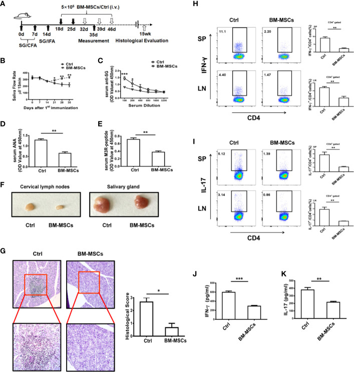
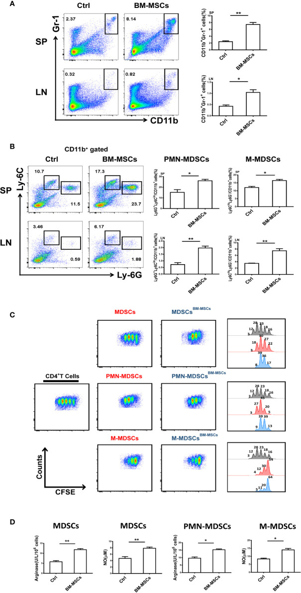
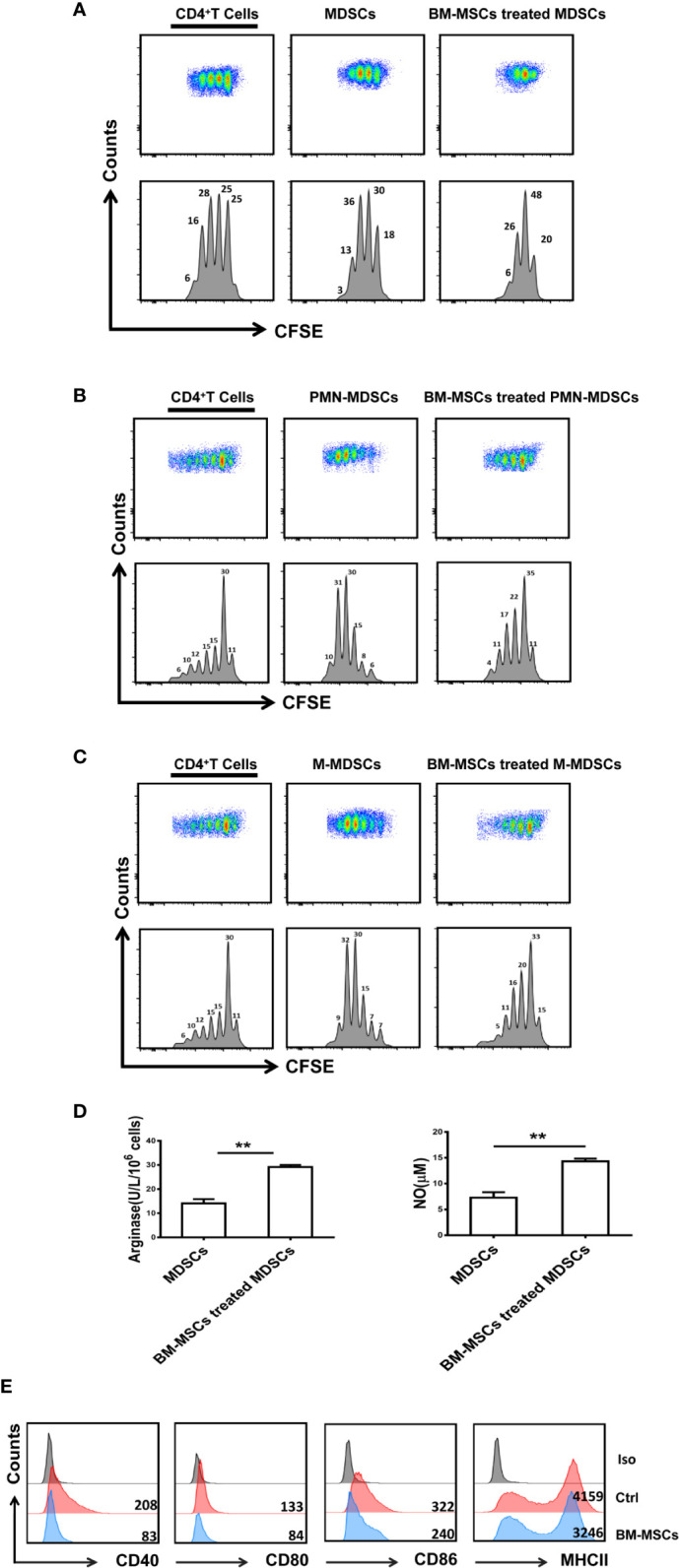
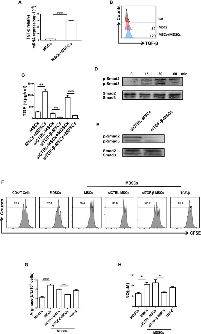
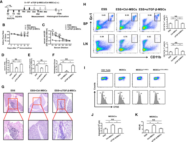
Primary Sjögren's syndrome (pSS) is a progressive systemic autoimmune disease characterized by lymphocytic infiltrates in exocrine glands, leading to the injury of salivary and lachrymal glands. Mesenchymal stem cells (MSCs) have been demonstrated to exert great potential in the treatment of various autoimmune diseases. Although MSCs have provide an effective therapeutic approach for SS treatment, the underlying mechanisms are still elusive. Our previous study has shown the reduced suppressive capacity of myeloid-derived suppressor cells (MDSCs) advanced the progression of experimental Sjögren's syndrome (ESS). In this study, we found that BM-MSCs significantly enhanced the suppressive function of MDSCs with high levels of Arginase and NO, decreased the levels of CD40, CD80, CD86, and MHC-II expression on MDSCs, thus attenuating the disease progression in ESS mice. Furthermore, the enhanced suppressive function of MDSCs was mediated by BM-MSC-secreted TGF-β, and the therapeutic effect of BM-MSCs in inhibiting ESS was almost abolished after silencing TGF-β in BM-MSCs. Taken together, our results demonstrated that BM-MSCs alleviated the ESS progression by up-regulating the immunosuppressive effect of MDSCs through TGF-β/Smad pathway, offering a novel mechanism for MSCs in the treatment of pSS.

Keywords: Sjögren’s syndrome; TGF-β; autoimmune disease; bone marrow-mesenchymal stem cell; myeloid-derived suppressor cell.


Conflict of interest statement

The authors declare that the research was conducted in the absence of any commercial or financial relationships that could be construed as a potential conflict of interest.

Figures

Figure 1
Figure 1
BM-MSCs suppress the progression of ESS. (A) Graphic scheme of ESS induction and BM-MSCs administration. C57BL/6 mice were immunized with SG/CFA on days 0 and 7, and mice were boosted with SG/IFA on day 14. Treatment groups were intravenously injected with 5×10⁵ BM-MSCs on days 18 and 25. Mice were sacrificed on day 35 (n=6). (B) The saliva flow rates were measured in each group. (C–E) Autoantibodies against SG antigens (C), ANA (D), and anti-M3R antibodies (E) were detected in the serum of ESS mice on day 35. (F) Representative graphs show the sizes of CLN and SG. (G) ESS mice were transferred with BM-MSCs on days 18, 25, 32, 39 and 46, the histological evaluation of glandular destruction in each group was performed on tissue sections of submandibular glands with H&E staining 15 weeks post first immunization. (H, I) Both proportions and numbers of CD4+IFN-γ+ Th1 cells (H) and CD4+IL-17+ Th17 cells (I) were measured in SP and CLN of mice with different treatment on day 35. (J, K) Serum levels of IFN-γ and IL-17 were detected in different groups on day 35. Data are shown as mean ± SD of three independent experiments, n=6/group. ***p < 0.001, **p < 0.01, *p < 0.05.
Figure 2
Figure 2
BM-MSCs enhance the suppressive capacity of MDSCs in ESS mice. (A, B) Proportions of CD11b+Gr-1+ MDSCs (A), M-MDSCs and PMN-MDSCs (B) were detected in SP and LN after BM-MSCs treatment (n=6). (C) Total MDSCs and their subsets from BM-MSCs treated group were isolated, and then co-cultured with CD4+T cells in the presence of anti-CD3 and anti-CD28 mAbs for 72 h (MDSC:T cell ratio 1:1). CD4+ T cell proliferation was evaluated by staining with CFSE. (D) The activity of arginase and the level of NO were measured in MDSCs and their subsets (n=6). Data are shown as means ± SD from three independent experiments, n=6/group. **p < 0.01, *p < 0.05.
Figure 3
Figure 3
BM-MSCs up-regulate the immunosuppressive function of MDSCs in vitro. (A–C) Total MDSCs (A), PMN-MDSCs (B), and M-MDSCs (C) isolated from the spleens of ESS mice were treated with BM-MSCs for 48 h, and then MDSCs were collected for co-culture with CD4+T cells in the presence of anti-CD3 and anti-CD28 mAbs for 72 h (MDSC:T cell ratio 1:1). CD4+ T cell proliferation was evaluated by staining with CFSE. (D) BM-MSCs treated MDSCs were used to measure the activity of arginase activity and the level of NO. (E) The expression of CD40, CD80, CD86, and MHCII on MDSCs in two groups was analyzed by flow cytometry. Data are shown as mean ± SD from three independent experiments. **p < 0.01.
Figure 4
Figure 4
The suppressive capacity of MDSCs was enhanced by BM-MSCs-secreted TGF-β. (A, B) The mRNA level (A) and protein level (B) of TGF-β in BM-MSCs co-cultured with MDSCs were analyzed by qRT-PCR and flow cytometry respectively. (C) The concentration of TGF-β in the conditioned medium of control MSCs and TGF-β silenced BM-MSCs with or without MDSCs were measured by ELISA. (D, E) The expression of phosphorylated Smad2/3 in MDSCs co-cultured with MSCs or TGF-β-silenced MSCs was determined by western blot. (F) TGF-β-silenced MSCs were co-cultured with MDSCs for 48 h, then MDSCs were collected to co-culture with CD4+T cells in the presence of anti-CD3 and anti-CD28 mAbs for 72 h (MDSC:T cell ratio 1:1). CD4+ T cell proliferation was evaluated by staining with CFSE. Recombinant mouse TGF-β (0.8ng/ml) was used as a control. (G, H) The activity of arginase activity and the level of NO were detected in each group. The Data are shown as mean ± SD from three independent experiments. ***p < 0.001, **p < 0.01, *p < 0.05. NS, no significance.
Figure 5
Figure 5
Knocking down TGF-β in BM-MSCs impairs their capability in inhibiting ESS development. (A) Graphic scheme of ESS induction and MSCs treatment. BM-MSCs transfected with TGF-β siRNA (siTGF-β) or negative control for 24 h, and then 5×10⁵ siTGF-β-MSCs or Ctrl-MSCs were intravenously injected on days 18 and 25 after the first immunization. Mice were sacrificed on day 35 (n=6). (B) The saliva flow rates were observed in each group. (C–E) Autoantibodies against SG antigens (C), ANA (D), and anti-M3R antibodies (E) were analyzed in the serum of mice with different treatment on day 35. (F, G) ESS mice were transferred with different BM-MSCs days 18, 25, 32, 39, and 46, the histological evaluation of glandular destruction in each group was performed on tissue sections of submandibular glands with H&E staining 15 weeks post first immunization. (H) Percentages of MDSCs in spleen and LNs were measured in each group on day 35. (I) MDSCs from different groups were isolated on day 35, and then co-cultured with CD4+T cells in the presence of anti-CD3 and anti-CD28 mAbs for 72 h (MDSC:T cell ratio 1:1). CD4+ T cell proliferation was evaluated by staining with CFSE. (J, K) The activity of arginase activity and the level of NO were detected in each group on day 35. Data are shown as mean± SD of three independent experiments, n=6/group. ***/###p < 0.001, **/##p < 0.01, */#p < 0.05, NS, no significance. * represents Ctrl-MSCs vs. siTGF-β-MSCs, #represents Ctrl-MSCs vs. ESS.

References

    1. Fox RI. Sjögren syndrome. Lancet (2005) 366(9482):321–31. 10.1016/S0140-6736(05)66990-5 - DOI - PubMed
    1. Fox PC. Autoimmune diseases and Sjögren syndrome: an autoimmune exocrinopathy. Ann N Y Acad Sci (2007) 1098:15–21. 10.1196/annals.1384.003 - DOI - PubMed
    1. Nocturne G, Mariette X. Advances in understanding the pathogenesis of primary Sjögren syndrome. Nat Rev Rheumatol (2013) 9(9):544–56. 10.1038/nrrheum.2013.110 - DOI - PubMed
    1. Lin X, Rui K, Deng J, Tian J, Wang X, Wang S, et al. Th17 cells play a critical role in the development of experimental Sjögren’s syndrome. Ann Rheum Dis (2015) 74(6):1302–10. 10.1136/annrheumdis-2013-204584 - DOI - PubMed
    1. Saito M, Otsuka K, Ushio A, Yamada A, Arakaki R, Kudo Y, et al. Unique Phenotypes and Functions of Follicular Helper T Cell and Regulatory T Cell in Sjögren’s Syndrome. Curr Rheumatol Rev (2018) 14(3):239–45. 10.2174/1573397113666170125122858 - DOI - PMC - PubMed

Publication types

MeSH terms