. 2020 Dec 22:11:606518.
doi: 10.3389/fimmu.2020.606518. eCollection 2020.

A Novel Gene Delivery Vector of Agonistic Anti-Radioprotective 105 Expressed on Cell Membranes Shows Adjuvant Effect for DNA Immunization Against Influenza

Affiliations

A Novel Gene Delivery Vector of Agonistic Anti-Radioprotective 105 Expressed on Cell Membranes Shows Adjuvant Effect for DNA Immunization Against Influenza

Tatsuya Yamazaki et al. Front Immunol. .

Abstract

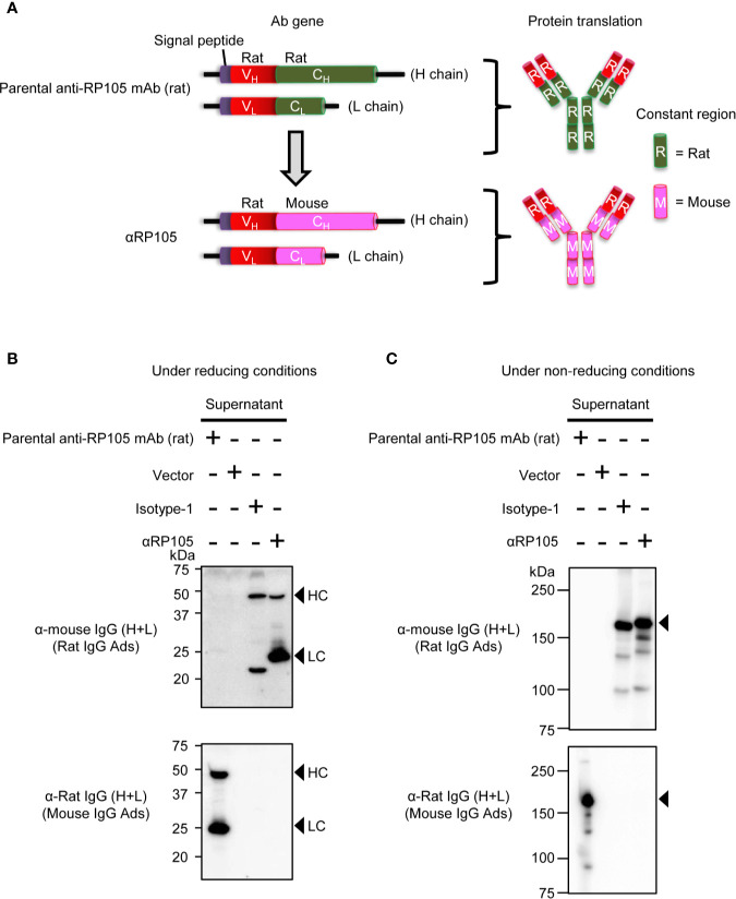
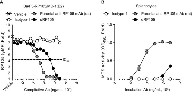
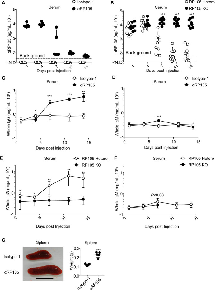
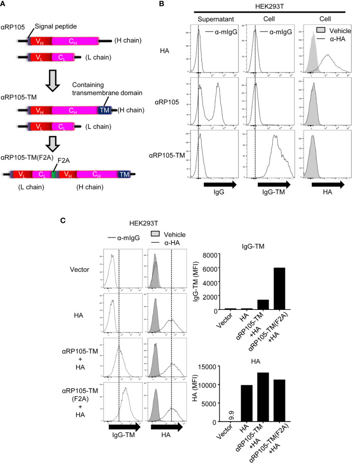
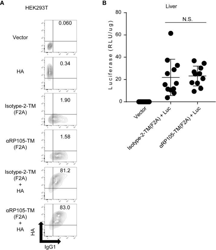
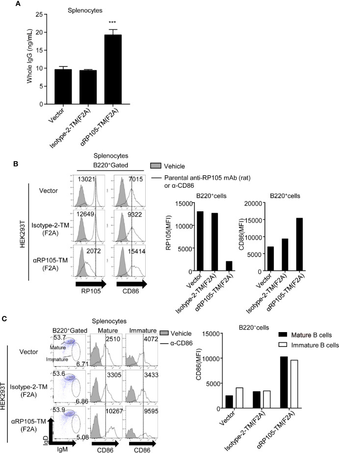
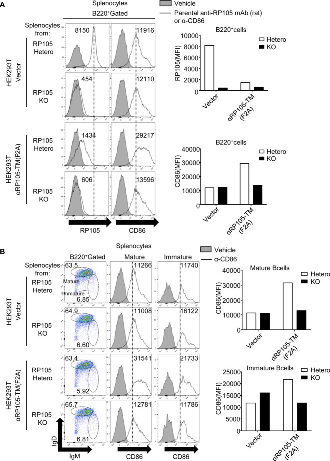
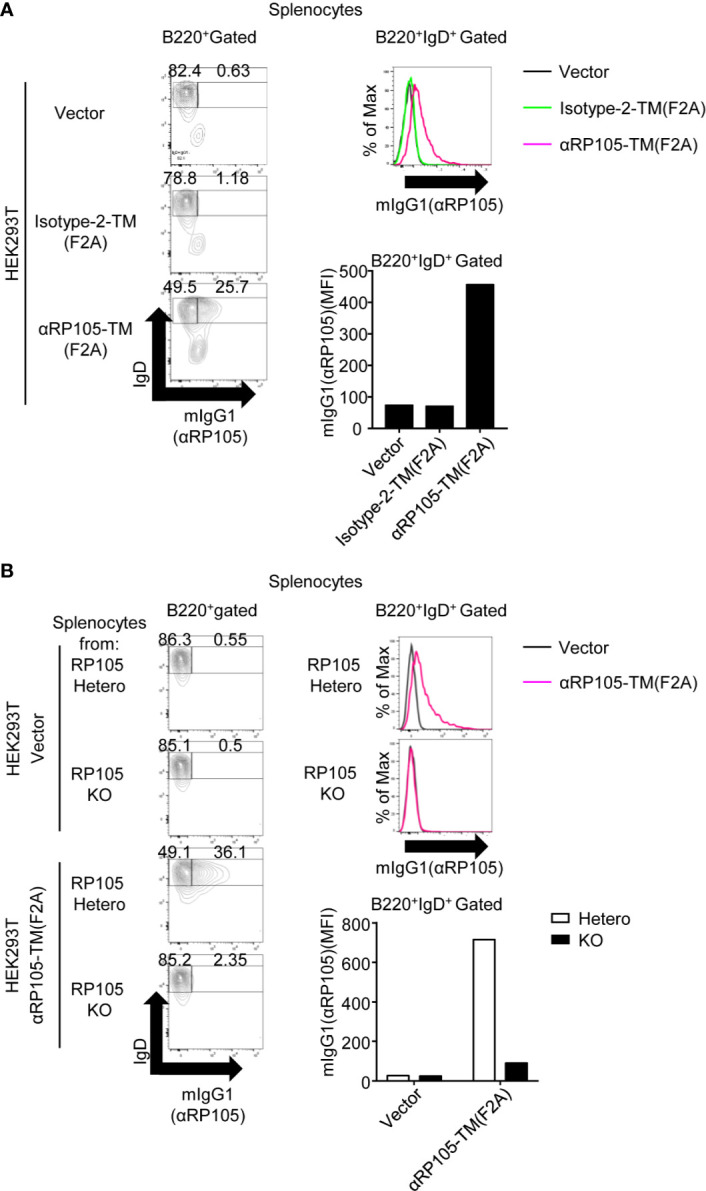
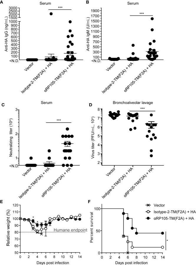
Radioprotective 105 (RP105) (also termed CD180) is an orphan and unconventional Toll-like receptor (TLR) that lacks an intracellular signaling domain. The agonistic anti-RP105 monoclonal antibody (mAb) can cross-link RP105 on B cells, resulting in the proliferation and activation of B cells. Anti-RP105 mAb also has a potent adjuvant effect, providing higher levels of antigen-specific antibodies compared to alum. However, adjuvanticity is required for the covalent link between anti-RP105 mAb and the antigen. This is a possible obstacle to immunization due to the link between anti-RP105 mAb and some antigens, especially multi-transmembrane proteins. We have previously succeeded in inducing rapid and potent recombinant mAbs in mice using antibody gene-based delivery. To simplify the covalent link between anti-RP105 mAb and antigens, we generated genetic constructs of recombinant anti-RP105 mAb (αRP105) bound to the transmembrane domain of the IgG-B cell receptor (TM) (αRP105-TM), which could enable the anti-RP105 mAb to link the antigen via the cell membrane. We confirmed the expression of αRP105-TM and the antigen hemagglutinin, which is a membrane protein of the influenza virus, on the same cell. We also found that αRP105-TM could activate splenic B cells, including both mature and immature cells, depending on the cell surface RP105 in vitro. To evaluate the adjuvanticity of αRP105-TM, we conducted DNA immunization in mice with the plasmids encoding αRP105-TM and hemagglutinin, followed by challenge with an infection of a lethal dose of an influenza virus. We then obtained partially but significantly hemagglutinin-specific antibodies and observed protective effects against a lethal dose of influenza virus infection. The current αRP105-TM might provide adjuvanticity for a vaccine via a simple preparation of the expression plasmids encoding αRP105-TM and of that encoding the target antigen.

Keywords: DNA immunization; RP105; adjuvant; agonistic antibody; antibody gene-vector delivery; cell membrane; influenza; targeting antigen to B cells.


Conflict of interest statement

The authors declare that the research was conducted in the absence of any commercial or financial relationships that could be construed as a potential conflict of interest.

Figures

Figure 1
Figure 1
In vitro expression of recombinant anti-RP105 (αRP105) from the Ab gene encoding the variable region (V) of anti-RP105 mAb combined with the constant region (C) in mice. (A) The genetic construction of recombinant anti-RP105 mAb (αRP105). (B, C) HEK293T cells were transfected with pCADEST1-empty (Vector), pCADEST1-anti-HA mIgG1 and pCADEST1-anti-HA mkappa (Isotyope-1), or pCADEST1-anti-RP105 mIgG1 and pCADEST1-anti-RP105 mkappa (αRP105). The supernatants were collected after 7 days. The supernatants and parental anti-RP105 mAb (rat) were detected by western blotting under reducing (B) or non-reducing (C) conditions, followed by probing with HRP-conjugated goat anti-mouse IgG adsorbed rat IgG or HRP-conjugated goat anti-rat IgG adsorbed mouse IgG. The indicated data are representative of two independent experiments.
Figure 2
Figure 2
αRP105 can also induce proliferation of splenocytes, but the activity is decreased. (A) Ba/F3 cells expressing RP105/MD-1 (B2) were incubated with the parental anti-RP105 mAb (rat), or the supernatant obtained from HEK293T cells expressing anti-HA (Isotype-1) or αRP105 after dilution from 2.5 × 10³ ng/ml, as indicated. Then, the cells were incubated with biotinylated parental anti-RP105 mAb (rat), followed by incubation with APC-conjugated streptavidin. Data were obtained using a BD LSRFortessa. Dose-dependent inhibitions are indicated as fold change normalized to geometric mean fluorescence (gMFI) from the incubation with 2.5 × 10³ ng/ml of parental anti-RP105 mAb (rat). The IC50 was determined by the average of the gMFI obtained from Isotype-1. (B) Splenocytes obtained from BALB/c mice were incubated with parental anti-RP105 mAb (rat), Isotype-1, or αRP105 after dilution from 2.5 × 10² ng/ml, as indicated, for 3 days. The proliferation and viability were assessed by an MTS assay. Data are indicated as the mean ± S.D. as fold change normalized to the absorbance at 490 nm from the incubation with 2.5 × 10² ng/ml of parental anti-RP105 mAb (rat). All indicated data are representative of at least two independent experiments.
Figure 3
Figure 3
αRP105 enhances the level of whole IgG in the serum, which depends on RP105, and enlarges the spleen. (A) BALB/c mice (n = 5) were subjected to HD with pCADEST1-anti-HA mIgG1 and pCADEST1-anti-HA mkappa (Isotyope-1) or pCADEST1-anti-RP105 mIgG1 and pCADEST1-anti-RP105 mkappa (αRP105). After the indicated times, the serum was obtained. The expression level of αRP105 was quantified using Ba/F3 cells expressing RP105/MD-1 (B2) using a BD LSRFortessa. The background level [38.1 (=101.581) ng/ml] was also determined using Ba/F3-null cells. (B) BALB/c RP105-Hetero or RP105-KO mice were subjected to HD with pCADEST1-anti-RP105 mIgG1 and pCADEST1-anti-RP105 mkappa. After the indicated times, the serum was obtained. The expression level of αRP105 was quantified by flow cytometry using Ba/F3 cells expressing RP105/MD-1 (B2). The background level [68.4 (=101.835) ng/ml] was also determined using Ba/F3-null cells. (C, D) Whole IgG (C) and IgM (D) levels in the serum (A) were analyzed by quantitative ELISA. (E, F) Whole IgG (E) and IgM (F) levels in the serum (B) were analyzed by quantitative ELISA. (G) Four days post-HD with the plasmids encoding Isotyope-1 or αRP105 into BALB/c WT mice (n = 5), the spleens were collected, and their weights were measured. The scale bar represents 1 cm. The indicated data (A, C, D, G) are representative of at least two independent experiments and are indicated as the mean ± S.D. The indicated data (B, E, F) are combined from two independent experiments (n = 8–9) and are indicated as the mean ± S.D. The detection limit was over 0.0147 (=10-1.832) mg/ml (C, E) or 0.0234 (=10-1.63) mg/ml (D, F). *P < 0.05, **P < 0.01, ***P < 0.001 (Student’s t-test).
Figure 4
Figure 4
The level of αRP105-TM expression on the cell membrane is enhanced using the F2A element. (A) The middle diagram indicates the genetic construction of anti-RP105 mIgG1 bound to the transmembrane (TM). The lower diagram indicates the genetic construction of anti-RP105 mIgG1-TM bound to anti-RP105 kappa via the F2A sequence. (B) HEK293T cells were transfected with pCADEST1-HA (A/PR8) as a control of membrane protein, pCADEST1-anti-RP105 mIgG1 and pCADEST1-anti-RP105 mkappa (αRP105), or pCADEST1-anti-RP105 mIgG1-TM and pCADEST1-anti-RP105 mkappa (αRP105-TM). Two days later, the supernatants and cells were collected. Ba/F3 cells expressing RP105/MD-1 (Balk) were incubated with the supernatants, followed by incubation with APC-conjugated anti-mouse IgG (Left panel). HEK293T cells were also incubated with APC-conjugated anti-mouse IgG (Middle panel) or biotinylated anti-HA IgG1, followed by incubation with APC-conjugated streptavidin (Right panel). The expression level was analyzed using a BD FACSCanto II. (C) HEK293T cells were transfected with pCADEST1-empty (Vector), pCADEST1-HA (A/PR8), and pCADEST1-anti-RP105 mIgG1-TM and pCADEST1-anti-RP105 mkappa (αRP105-TM) or pCADEST1-anti-RP105 kappa-F2A-anti-RP105 mIgG1-TM [αRP105-TM (F2A)]. Two days later, the cells were collected and incubated with APC-conjugated anti-mouse IgG (Left panel) or biotinylated anti-HA IgG1, followed by incubation with APC-conjugated streptavidin (Right panel). The expression level was analyzed using a BD LSRFortessa. All indicated data are representative of at least two independent experiments.
Figure 5
Figure 5
Antigen expression level in vivo and in vitro is not significantly different with co-expression of Isotype-2-TM and αRP105-TM. (A) HEK293T cells were co-transfected as indicated. Isotype-2-TM (F2A) was expressed from pCADEST1-anti-OVA kappa-F2A-anti-OVA mIgG1-TM ( Figure S4 ). Two days later, these cells were collected and incubated with FITC-conjugated anti-mouse IgG1 and anti-HA IgD, followed by incubation with APC-conjugated anti-mouse IgD. The indicated numbers represent the ratio (%) of IgG1+HA+ cells in total. The expression level was analyzed using a BD LSRFortessa. The indicated data are representative of at least two independent experiments. (B) Female BALB/c mice were subjected to HD with either pCADEST1-empty (Vector), pCADEST1-anti-OVA kappa-F2A-anti-OVA mIgG1-TM [Isotype-2-TM (F2A)] and pCADEST1-luciferase (Luc), or pCADEST1-anti-RP105 kappa-F2A-anti-RP105 mIgG1-TM [αRP105-TM (F2A)] and pCADEST1-Luc, as indicated. One day later, the liver was obtained, and luciferase activity was determined. The indicated data are combined from three independent experiments (n = 11–12) and indicated as the mean ± S.D. N.S., not significant (Mann-Whitney test).
Figure 6
Figure 6
αRP105-TM can activate both mature and immature B cells. (A) HEK293T cells were transfected with pCADEST1-empty (Vector), pCADEST1-anti-OVA kappa-F2A-anti-OVA mIgG1/TM [Isotype-2-TM (F2A)], or pCADEST1-anti-RP105 kappa-F2A-anti-RP105 mIgG1-TM [αRP105-TM (F2A)]. Two days later, the splenocytes obtained from BALB/c mice were incubated with the HEK293T cells for 7 days. The supernatant was obtained, and the whole IgG level was measured by quantitative ELISA. The detection limit was over 3.91 ng/ml. ***P < 0.001 (One-way ANOVA). (B, C) HEK293T cells were transfected as indicated. Two days later, splenocytes obtained from BALB/c mice were co-cultured with HEK293T cells for 2 days. Then, the splenocytes, which were gated on B220 (B), IgMlow IgDhigh as mature B cells (Middle panel in C), or IgMhigh IgDlow as immature B cells (Right panel in C), were also incubated with biotinylated parental anti-RP105 mAb (rat) or anti-CD86 antibodies as indicated, followed by incubation with PE-conjugated streptavidin. The indicated numbers on each gate (Left panel in C) respectively represent the percentage of cells of mature or immature B cells gated on B220+ cells. The numbers in the histogram represent MFI. All indicated data are representative of two independent experiments.
Figure 7
Figure 7
αRP105-TM can activate both immature and mature B cells depending on RP105. (A) HEK293T cells were transfected with pCADEST1-empty (Vector) or pCADEST1-anti-RP105 kappa-F2A-anti-RP105 mIgG1-TM [αRP105-TM (F2A)]. Two days later, the splenocytes that were obtained from RP105-Hetero and RP105-KO BALB/c mice were co-cultured with HEK293T cells for 2 days. The splenocytes, which were gated on B220, were also incubated with biotinylated parental anti-RP105 mAb (rat) or anti-CD86 antibodies as indicated, followed by incubation with PE-conjugated streptavidin. (B) The splenocytes, which were gated on IgMlow IgDhigh as mature B cells (Middle panel) or IgMhigh IgDlow as immature B cells (Right panel), were also incubated with biotinylated anti-CD86 antibodies, followed by incubation with PE-conjugated streptavidin. The indicated numbers on each gate (Left panel in B) respectively represent the percentage of cells of mature or immature B cells gated on B220+ cells. The numbers in the histogram represent MFI. The expression level was analyzed using a BD LSRFortessa. All indicated data are representative of two independent experiments.
Figure 8
Figure 8
αRP105-TM can associate with the B cell membrane, depending on RP105. (A) From Figures 6B, C , the splenocytes were collected and incubated with FITC-conjugated anti-mouse IgG1 and APC-conjugated anti-mouse IgD. (B) From Figures 7A, B , the splenocytes were collected and incubated with FITC-conjugated anti-mouse IgG1 and APC-conjugated anti-mouse IgD. The indicated numbers on each gate in the left panel represents the percentage of cells of IgD+mIgG1- or IgD+mIgG1+ gated on B220+ cells, respectively. The histogram and bar graph in the panels on the right represent the level of mIgG1, which represents the level of αRP105 on B220+ IgD+cells. The expression level was analyzed using a BD LSRFortessa. All indicated data are representative of two independent experiments.
Figure 9
Figure 9
αRP105-TM can significantly increase the level of neutralizing antibodies against the influenza virus and provide protection. (A–F) Female BALB/c mice were subjected to HD with either pDEST1-empty (Vector), pCADEST1-anti-OVA kappa-F2A-anti-OVA mIgG1-TM [Isotype-2-TM (F2A)] and pCADEST1-HA (A/PR8), or pCADEST1-anti-RP105 kappa-F2A-anti-RP105 mIgG1-TM [αRP105-TM (F2A)] and pCADEST1-HA (A/PR8). Fourteen days later, the serum was obtained, and the level of anti-HA IgG (A) and that of anti-HA IgM (B) were measured by quantitative ELISA. The detection limit was over 1.2 ng/ml (A) or 15.6 U/ml (B). (C) The neutralizing titer against the influenza virus was also determined by a micro-neutralization assay. The detection limit was over 10. (D) The next day, the mice were infected with a lethal dose of A/PR8 virus (1,000 PFU). Three days post-infection, the bronchoalveolar lavage specimens were obtained, and viral titers were determined by a plaque assay. (E, F) The body weight (E) and survival rates (F) in another group were monitored for 14 days. The body weight was expressed relative to the initial mean body weight of each group. (A–D) The indicated data are combined either from four independent experiments (n = 27–29) (A), three independent experiments (n = 21–22) (B), two independent experiments (n = 17–18) (C), or two independent experiments (n = 13–14) (D). (E, F) The indicated data are representative of three independent experiments. All error bars represent the S.E.M. *P < 0.05, ***P < 0.001 [(AE), Mann-Whitney test; (F), Log-rank test].

Similar articles

Cited by

References

    1. Carter PJ, Lazar GA. Next generation antibody drugs: pursuit of the ‘high-hanging fruit’. Nat Rev Drug Discovery (2018) 17(3):197–223. 10.1038/nrd.2017.227 - DOI - PubMed
    1. Qin S, Xu L, Yi M, Yu S, Wu K, Luo S. Novel immune checkpoint targets: moving beyond PD-1 and CTLA-4. Mol Cancer (2019) 18(1):155. 10.1186/s12943-019-1091-2 - DOI - PMC - PubMed
    1. Nunes-Alves C. Blood is a very unusual fluid. Nat Immunol (2016) 17(1):S5–S. 10.1038/ni.3600 - DOI
    1. Chaplin JW, Chappell CP, Clark EA. Targeting antigens to CD180 rapidly induces antigen-specific IgG, affinity maturation, and immunological memory. J Exp Med (2013) 210(10):2135–46. 10.1084/jem.20130188 - DOI - PMC - PubMed
    1. Erdő-Bonyár S, Rapp J, Minier T, Ráth G, Najbauer J, Czirják L, et al. Toll-Like Receptor Mediated Activation of Natural Autoantibody Producing B Cell Subpopulations in an Autoimmune Disease Model. Int J Mol Sci (2019) 20(24):6152. 10.3390/ijms20246152 - DOI - PMC - PubMed

Publication types

MeSH terms