

Overexpression of LMP-1 Decreases Apoptosis in Human Nucleus Pulposus Cells via Suppressing the NF- κ B Signaling Pathway
- PMID: 33414896
- PMCID: PMC7752285
- DOI: 10.1155/2020/8189706
Overexpression of LMP-1 Decreases Apoptosis in Human Nucleus Pulposus Cells via Suppressing the NF- κ B Signaling Pathway
Abstract
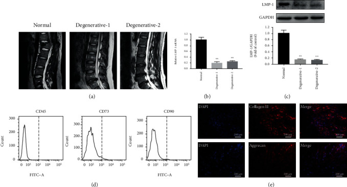
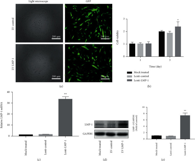
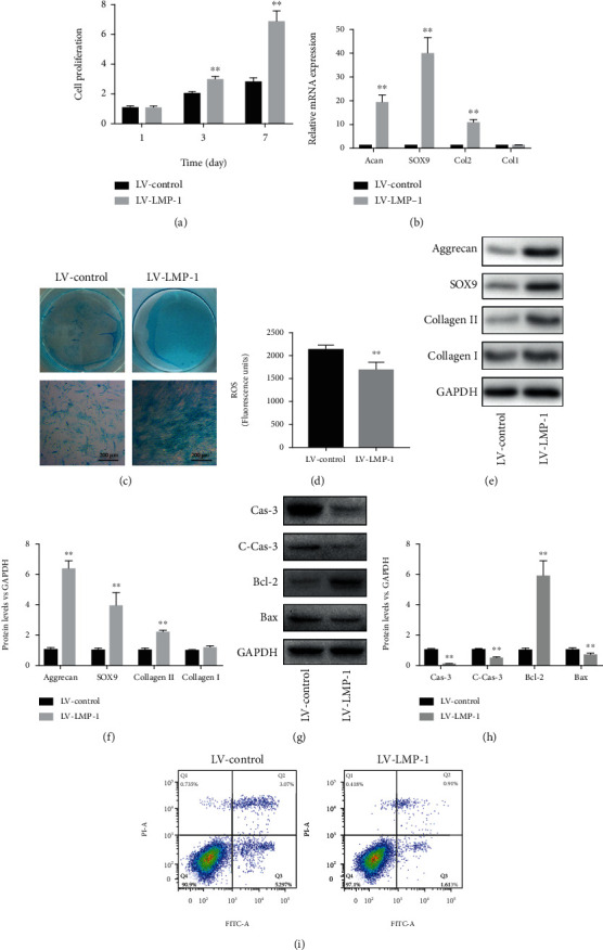
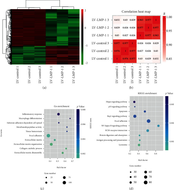
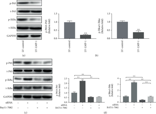
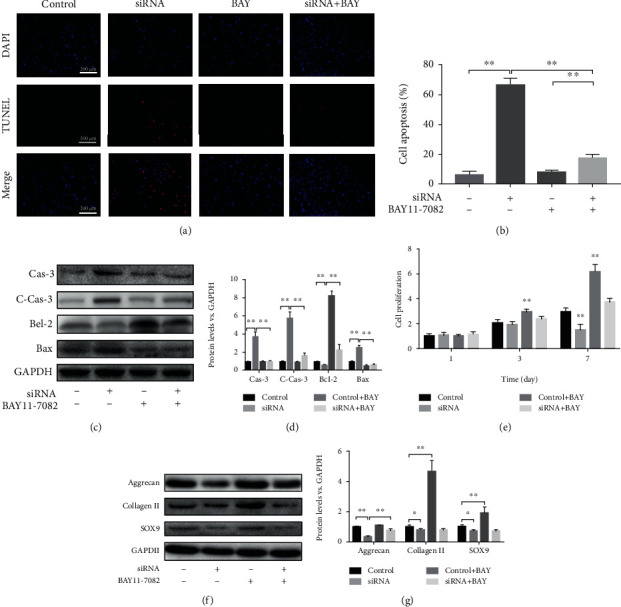
Intervertebral disc degeneration (IDD) is a prevalent disease characterized by low back pain. Increasing extracellular matrix (ECM) synthesis and decreasing nucleus pulposus cell (NPC) apoptosis are promising strategies to recover degenerated NP. LIM mineralization protein- (LMP-) 1 has anti-inflammatory potential and is a promising gene target for the treatment of NP degeneration. In this study, we measured the expression of LMP-1 in the NP of patients. Then, we constructed LMP-1-overexpressing NPCs using lentiviral vectors and investigated the effects of LMP-1 on cell proliferation, apoptosis, and ECM synthesis in NPCs. The results showed that LMP-1 was highly expressed in the NP of patients. LMP-1 overexpression significantly increased proliferation and decreased apoptosis in NPCs. The expression of collagen II and sulfated glycosaminoglycan (sGAG) in NPCs was also upregulated after LMP-1 was overexpressed. Moreover, we demonstrated that LMP-1 decreased apoptosis of NPCs by inhibiting NF-κB signaling activation. These findings suggest that LMP-1 plays an essential role in mediating apoptosis in NPCs by regulating NF-κB signaling and can be used as a gene target for the treatment of IDD.
Copyright © 2020 Yuan Liu et al.
Conflict of interest statement
The authors declare that they have no conflicts of interest.
Figures

Similar articles
-
LIM mineralization protein-1 suppresses TNF-α induced intervertebral disc degeneration by maintaining nucleus pulposus extracellular matrix production and inhibiting matrix metalloproteinases expression.J Orthop Res. 2015 Mar;33(3):294-303. doi: 10.1002/jor.22732. Epub 2014 Oct 21. J Orthop Res. 2015. PMID: 25336289
-
Hemeoxygenase-1 Suppresses IL-1β-Induced Apoptosis Through the NF-κB Pathway in Human Degenerative Nucleus Pulposus Cells.Cell Physiol Biochem. 2018;46(2):644-653. doi: 10.1159/000488632. Epub 2018 Mar 28. Cell Physiol Biochem. 2018. PMID: 29617687
-
A20 regulates inflammation through autophagy mediated by NF-κB pathway in human nucleus pulposus cells and ameliorates disc degeneration in vivo.Biochem Biophys Res Commun. 2021 Apr 16;549:179-186. doi: 10.1016/j.bbrc.2021.02.115. Epub 2021 Mar 4. Biochem Biophys Res Commun. 2021. PMID: 33677390
-
DBC1 promotes intervertebral disc degeneration by activating NF-κB pathway and inhibiting SIRT1 activity.Life Sci. 2025 Jul 15;373:123689. doi: 10.1016/j.lfs.2025.123689. Epub 2025 May 6. Life Sci. 2025. PMID: 40339956
-
NF-κB signalling pathways in nucleus pulposus cell function and intervertebral disc degeneration.Cell Prolif. 2021 Jul;54(7):e13057. doi: 10.1111/cpr.13057. Epub 2021 May 24. Cell Prolif. 2021. PMID: 34028920 Free PMC article. Review.
Cited by
-
Natural products can modulate inflammation in intervertebral disc degeneration.Front Pharmacol. 2023 Feb 16;14:1150835. doi: 10.3389/fphar.2023.1150835. eCollection 2023. Front Pharmacol. 2023. PMID: 36874009 Free PMC article. Review.
-
SIRT1 Attenuates Apoptosis of Nucleus Pulposus Cells by Targeting Interactions between LC3B and Fas under High-Magnitude Compression.Oxid Med Cell Longev. 2021 Dec 27;2021:2420969. doi: 10.1155/2021/2420969. eCollection 2021. Oxid Med Cell Longev. 2021. PMID: 34987698 Free PMC article.
-
Regulating the fate of stem cells for regenerating the intervertebral disc degeneration.World J Stem Cells. 2021 Dec 26;13(12):1881-1904. doi: 10.4252/wjsc.v13.i12.1881. World J Stem Cells. 2021. PMID: 35069988 Free PMC article. Review.
References
-
- Antoniou J., Steffen T., Nelson F., et al. The human lumbar intervertebral disc: evidence for changes in the biosynthesis and denaturation of the extracellular matrix with growth, maturation, ageing, and degeneration. The Journal of Clinical Investigation. 1996;98(4):996–1003. doi: 10.1172/JCI118884. - DOI - PMC - PubMed
MeSH terms
Substances
LinkOut - more resources
Full Text Sources
Miscellaneous
