. 2020 Dec 1;10(12):4234-4250.
eCollection 2020.

CD4+ T cell exhaustion leads to adoptive transfer therapy failure which can be prevented by immune checkpoint blockade

Affiliations

CD4+ T cell exhaustion leads to adoptive transfer therapy failure which can be prevented by immune checkpoint blockade

Jinfei Fu et al. Am J Cancer Res. .

Abstract

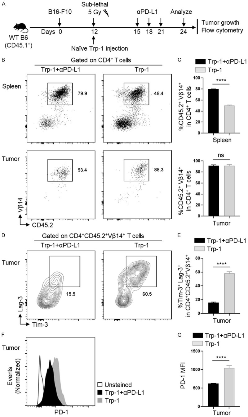
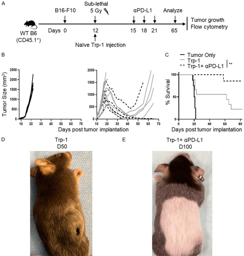
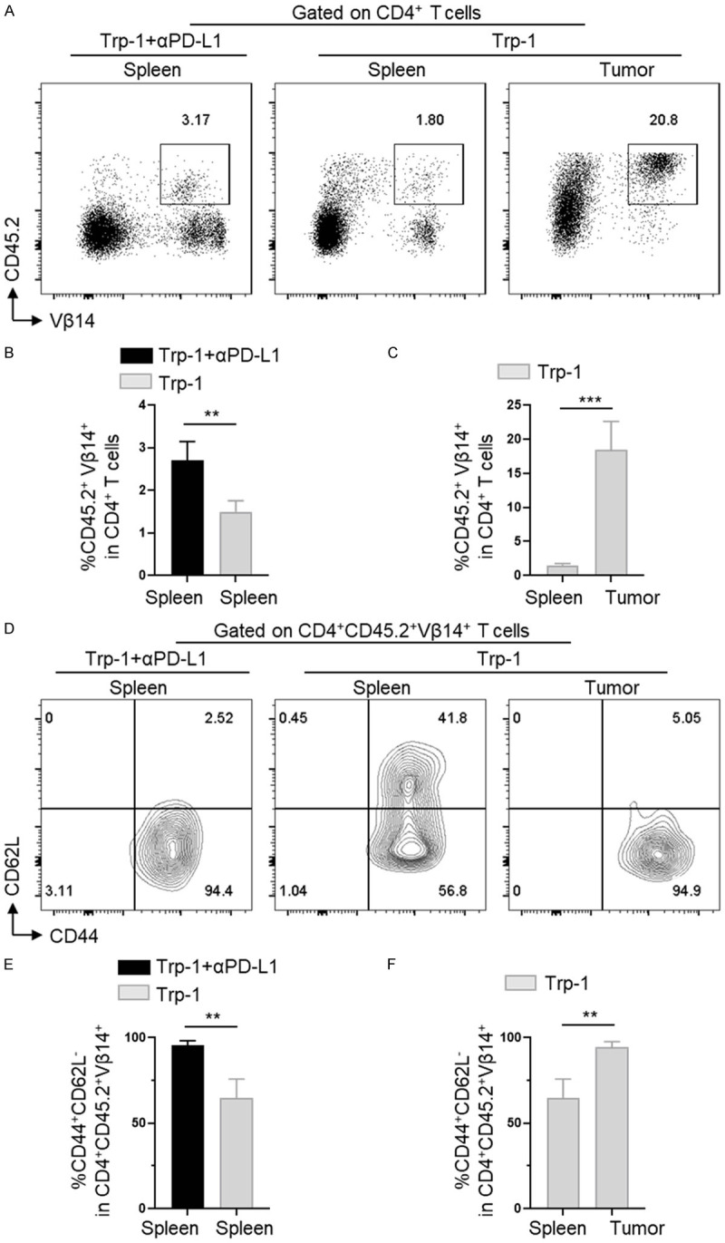
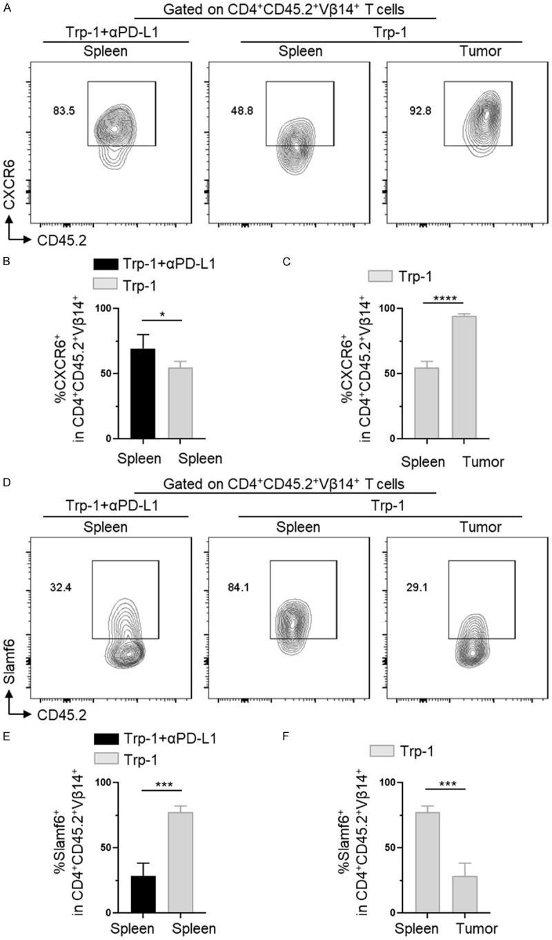
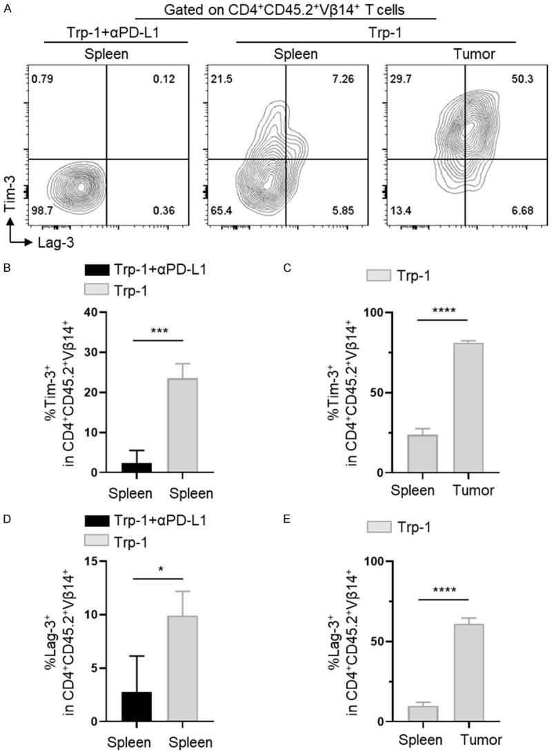
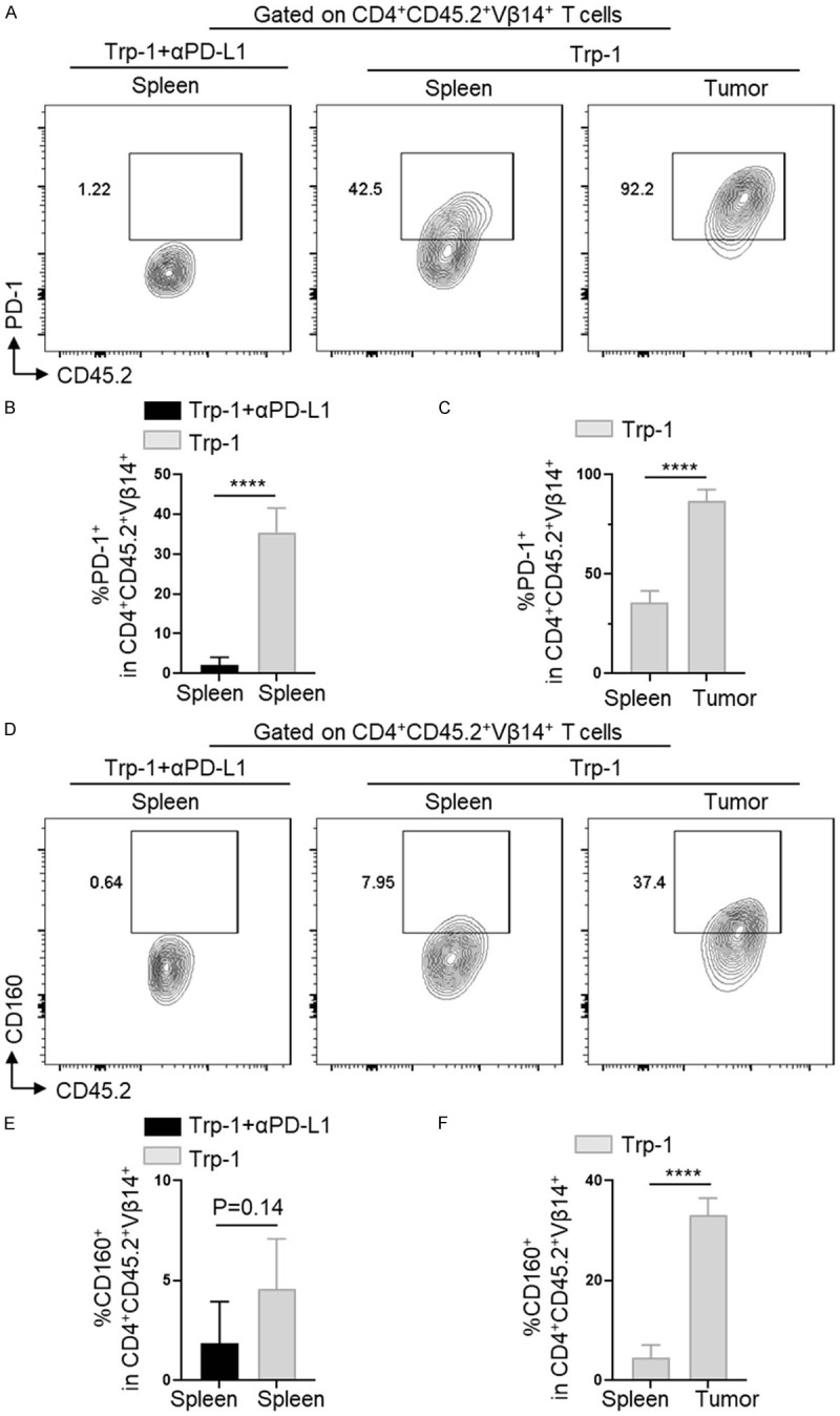
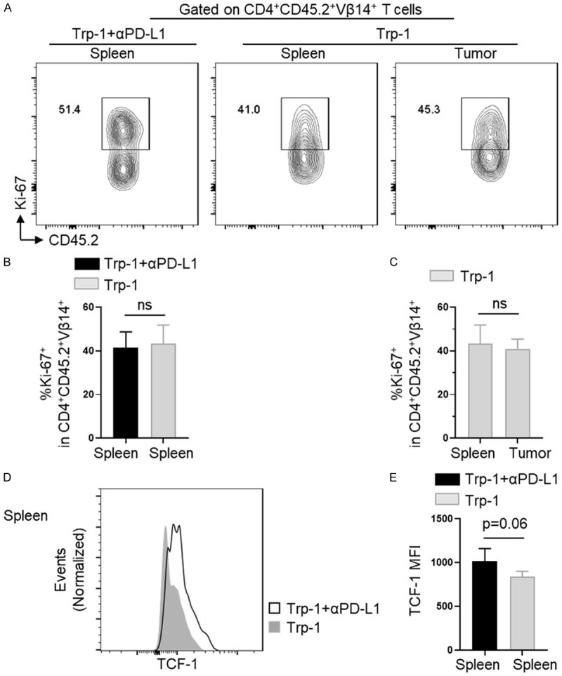
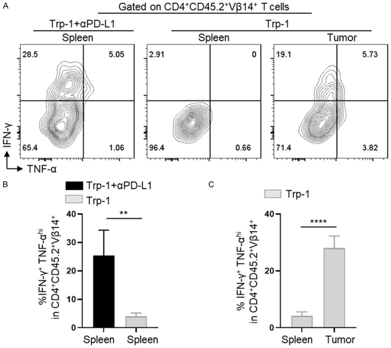
Cytotoxic CD8+ T cell exhaustion is one of the mechanisms underlying the tumor immune escape. The paradigm-shifting immune checkpoint therapy can mitigate CD8+ T lymphocyte exhaustion, reinvigorate the anticancer immunity, and achieve durable tumor regression for some patients. Emerging evidence indicates that CD4+ T lymphocytes also have a critical role in anticancer immunity, either by directly applying cytotoxicity toward cancer cells or as a helper to augment CD8+ T cell cytotoxicity. Whether anticancer CD4+ T lymphocytes undergo exhaustion during immunotherapy of solid tumors remains unknown. Here we report that melanoma antigen TRP-1/gp75-specific CD4+ T lymphocytes exhibit an exhaustion phenotype after being adoptively transferred into mice bearing large subcutaneous melanoma. Exhaustion of these CD4+ T lymphocytes is accompanied with reduced cytokine release and increased expression of inhibitory receptors, resulting in loss of tumor control. Importantly, we demonstrate that PD-L1 immune checkpoint blockade can prevent exhaustion, induce proliferation of the CD4+ T lymphocytes, and consequently prevent tumor recurrence. Therefore, when encountering an excessive amount of tumor antigens, tumor-reactive CD4+ T lymphocytes also enter the exhaustion state, which can be prevented by immune checkpoint blockade. Our results highlight the importance of tumor-specific CD4+ T lymphocytes in antitumor immunity and suggest that the current immune checkpoint blockade therapy may achieve durable anticancer efficacy by rejuvenating both tumor antigen-specific CD8+ T lymphocytes and CD4+ T lymphocytes.

Keywords: CD4+ T cells; adoptive transfer therapy; exhaustion; immune checkpoint blockade.


Conflict of interest statement

None.

Figures

Figure 1
Figure 1
Anticancer activity of adoptively transferred Trp-1 CD4+ T lymphocytes in mice bearing subcutaneous melanoma and efficacy of co-treatment with immune checkpoint blockade. A. Schematic diagram showing the treatment regimen and the timeline for mice bearing melanoma. On day 24 post-tumor inoculation, the adoptively transferred Trp-1 CD4+ T lymphocytes isolated from irradiated WT B6 (CD45.1+) recipient mice were examined. All plots and histograms of flow cytometric analysis were gated on adoptively transferred CD4+ T lymphocytes among living splenocytes and TILs. B and C. Proportions of Trp-1 CD4+ T lymphocytes (CD45.2+Vβ14+) among gated total CD4+ T lymphocytes isolated from spleens and tumors on day 24. D and E. Representative flow and bar graphs of Tim-3 and Lag-3 expression on tumor infiltrating Trp-1 CD4+ T lymphocytes of each treatment group. F and G. PD-1 expression in tumor infiltrating Trp-1 CD4+ T lymphocytes of each treatment group. Experiments were repeated three times independently and data are presented as mean ± SD (n=4) from one representative experiment. Not significant = ns: P value >0.05; *: P<0.05; **: P<0.01; ***: P<0.001; ****: P<0.0001. Unpaired 2-tailed Student’s t test.
Figure 2
Figure 2
Adoptively transferred CD4+ T lymphocytes induced temporary regression of large established tumors, and prevention of tumor relapse by immune checkpoint blockade. (A) Schematic diagram showing the treatment regimen and the timeline for mice bearing large melanoma. WT B6 (CD45.1+) mice bearing B16F10 mouse melanoma (tumor diameter ~0.8 cm) received sublethal 5 Gy whole body irradiation and iv injected with tumor-specific Trp-1 CD4+ T lymphocytes. On day 15, 18 and 21, αPD-L1 or PBS control was administered for three times. Tumor size was measured with caliper every two days. (B) The growth curves of subcutaneous B16F10 melanoma in mice receiving three different treatment regimens: no treatment, Trp-1 CD4+ T cells/PBS, and Trp-1 CD4+ T cells/αPD-L1. (C) Survival curves of mice receiving different treatment regimens based on the log-rank test. Data shown were combined from two independent experiments (n≥6 per group). **: P<0.01. The tumor pictures of representative recipient mice that received Trp-1 CD4+ T cells/PBS on day 50 (D) and Trp-1 CD4+ T cells/αPD-L1 on day 100 (E).
Figure 3
Figure 3
Checkpoint blockade induced proliferative response and terminal effector phenotype in Trp-1 CD4+ T lymphocytes. WT B6 (CD45.1+) mice bearing B16F10 murine melanoma were adoptively transferred with 5 × 10⁴ naïve Trp-1 CD4+ T lymphocytes. On day 65 post-tumor inoculation, Trp-1 CD4+ T lymphocytes were isolated from spleens and tumors and analyzed by flow cytometry. All plots and histograms were gated on transferred Trp-1 CD4+ T lymphocytes among living splenocytes and TILs. A-C. The proportions of Trp-1 CD4+ T lymphocytes (CD45.2+Vβ14+) among gated total CD4+ T lymphocytes in spleens (Trp-1 CD4+ T cells/αPD-L1) or in spleens and tumors (Trp-1 CD4+ T cells/PBS). D-F. The percentage of CD62L-CD44+ terminal effector T lymphocytes among the transferred Trp-1 CD4+ T lymphocytes. Data are presented as mean ± SD (n=4). **: P<0.01, ***: P<0.001. Unpaired 2-tailed Student’s t test.
Figure 4
Figure 4
Checkpoint blockade prevented tumor relapse and altered the expression of CXCR6 and Slamf6 in Trp-1 CD4+ T lymphocytes. WT B6 (CD45.1+) mice bearing B16F10 mouse melanoma were adoptively transferred with 5 × 10⁴ naïve Trp-1 cells. On day 65 post tumor inoculation, adoptively transferred Trp-1 CD4+ T lymphocytes were isolated from spleens and tumors and analyzed by flow cytometry. All plots and histograms of flow cytometric analysis were gated on adoptively transferred CD4+ T lymphocytes among living splenocytes and TILs. A-C. The percentage of CXCR6+ effector T lymphocytes among the transferred CD45.2+Vβ14+ Trp-1 cells in spleens (Trp-1 CD4+ T cells/αPD-L1) or in spleens and tumors (Trp-1 CD4+ T cells/PBS). D-F. Representative contour plots and bar graphs display the percentage of Slamf6+ T lymphocytes among the transferred CD45.2+Vβ14+ Trp-1 cells in spleens only (Trp-1 CD4+ T cell/αPD-L1) or spleens and tumors (Trp-1 CD4+ T cells/PBS). Data are presented as mean ± SD (n=4). *: P<0.05, ***: P<0.001, ****: P<0.0001. Unpaired 2-tailed Student’s t test.
Figure 5
Figure 5
Checkpoint blockade inhibited the expression of exhaustion markers Tim-3 and Lag-3 in Trp-1 CD4+ T lymphocytes. WT B6 (CD45.1+) mice bearing B16F10 mouse melanoma were adoptively transferred with 5 × 10⁴ naïve Trp-1 cells. Adoptively transferred Trp-1 cells isolated from spleens and tumors on day 65 post tumor inoculation and were analyzed by flow cytometry. All plots and histograms of flow cytometric analysis were gated on adoptively transferred CD4+ T lymphocytes among living splenocytes and TILs. A. The percentage of Tim-3+ and Lag-3+ exhausted T lymphocytes among the transferred CD45.2+Vβ14+ Trp-1 cells in spleens only (Trp-1 CD4+ T cells/αPD-L1) or in both spleens and tumors (Trp-1 CD4+ T cells/PBS). B and C. The percentage of Tim-3+ exhausted T lymphocytes among the transferred CD45.2+Vβ14+ Trp-1 cells in spleens only (Trp-1 CD4+ T cells/αPD-L1) or both spleens and tumors (Trp-1 CD4+ T cells/PBS). D and E. Bar graphs display the percentage of Lag-3+ exhausted T lymphocytes among the transferred CD45.2+Vβ14+ Trp-1 cells in spleens only (Trp-1 CD4+ T cells/αPD-L1) or both spleens and tumors (Trp-1 CD4+ T cells/PBS). Data are presented as mean ± SD (n=4). *: P<0.05, ***: P<0.001, ****: P<0.0001. Unpaired 2-tailed Student’s t test.
Figure 6
Figure 6
Checkpoint blockade inhibited the expression of exhaustion markers PD-1 and CD160 in Trp-1 CD4+ T lymphocytes. WT B6 (CD45.1+) mice bearing B16F10 murine melanoma were adoptively transferred with 5 × 10⁴ naïve Trp-1 cells. Adoptively transferred Trp-1 cells isolated from spleens and tumors on day 65 post-tumor inoculation and analyzed by flow cytometry. All plots and histograms of flow cytometric analysis were gated on adoptively transferred CD4+ T lymphocytes among living splenocytes and TILs. A-C. The percentage of PD-1+ T lymphocytes among the transferred CD45.2+Vβ14+ Trp-1 cells in spleens only (Trp-1 CD4+ T cells/αPD-L1) or both spleens and tumors (Trp-1 CD4+ T cells/PBS). D-F. Representative contour plots and bar graphs display the percentage of CD160+ T lymphocytes among the transferred CD45.2+Vβ14+ Trp-1 cells in spleens only (Trp-1 CD4+ T cells/αPD-L1) or both spleens and tumors (Trp-1 CD4+ T cells/PBS). Data are presented as mean ± SD (n=4). ****: P<0.0001. Unpaired 2-tailed Student’s t test.
Figure 7
Figure 7
Checkpoint blockade did not affect the expression of Ki-67 while induced a progenitor-effector T cell phenotype in peripheral lymphoid organs. WT B6 (CD45.1+) mice bearing B16F10 mouse melanoma were adoptively transferred with 5 × 10⁴ naïve Trp-1 CD4+ T lymphocytes. On day 65 post-tumor inoculation, adoptively transferred Trp-1 CD4+ T lymphocytes were isolated from spleens and tumors and were analyzed by flow cytometry. All plots and histograms of flow cytometric analysis were gated on adoptively transferred CD4+ T lymphocytes among living splenocytes and TILs. A-C. The percentage of Ki-67+ T lymphocytes among the transferred CD45.2+Vβ14+ Trp-1 cells in spleens only (Trp-1 CD4+ T cells/αPD-L1) or both spleens and tumors (Trp-1 CD4+ T cells/PBS). D and E. Overlay histogram and bar graphs display the mean fluoresce intensity of TCF-1 among the transferred CD45.2+Vβ14+ Trp-1 cells in spleens (Trp-1 CD4+ T cells/αPD-L1 group and Trp-1 CD4+ T cells/PBS group). Data are presented as mean ± SD (n=4). ns: P>0.05. Unpaired 2-tailed Student’s t test.
Figure 8
Figure 8
Checkpoint blockade increased Trp-1 CD4+ T lymphocyte production of anticancer cytokines. WT B6 (CD45.1+) mice bearing B16F10 mouse melanoma were adoptively transferred with 5 × 10⁴ naïve Trp-1 CD4+ T lymphocytes. On day 65 post-tumor inoculation, adoptively transferred Trp-1 cells were isolated from spleens and tumors and analyzed by flow cytometry. All plots and histograms of flow cytometric analysis were gated on adoptively transferred CD4+ T cells among living splenocytes and TILs. A-C. The percentage of IFN-γ+ TNF-αhi T lymphocytes among the transferred CD45.2+Vβ14+ Trp-1 cells in spleens only (Trp-1 CD4+ T cells/αPD-L1) or both spleens and tumors (Trp-1 CD4+ T cells/PBS). Data are presented as mean ± SD (n=4). **: P<0.01, ****: P<0.0001. Unpaired 2-tailed Student’s t test.

Similar articles

Cited by

References

    1. Wu J, Zhang H, Shi X, Xiao X, Fan Y, Minze LJ, Wang J, Ghobrial RM, Xia J, Sciammas R, Li XC, Chen W. Ablation of transcription factor IRF4 promotes transplant acceptance by driving allogenic CD4(+) T cell dysfunction. Immunity. 2017;47:1114–1128. e1116. - PMC - PubMed
    1. Zhang H, Wu J, Zou D, Xiao X, Yan H, Li XC, Chen W. Ablation of interferon regulatory factor 4 in T cells induces “memory” of transplant tolerance that is irreversible by immune checkpoint blockade. Am J Transplant. 2019;19:884–893. - PMC - PubMed
    1. Blank CU, Haining WN, Held W, Hogan PG, Kallies A, Lugli E, Lynn RC, Philip M, Rao A, Restifo NP, Schietinger A, Schumacher TN, Schwartzberg PL, Sharpe AH, Speiser DE, Wherry EJ, Youngblood BA, Zehn D. Defining ‘T cell exhaustion’. Nat Rev Immunol. 2019;19:665–674. - PMC - PubMed
    1. Sekine T, Perez-Potti A, Nguyen S, Gorin JB, Wu VH, Gostick E, Llewellyn-Lacey S, Hammer Q, Falck-Jones S, Vangeti S, Yu M, Smed-Sörensen A, Gaballa A, Uhlin M, Sandberg JK, Brander C, Nowak P, Goepfert PA, Price DA, Betts MR, Buggert M. TOX is expressed by exhausted and polyfunctional human effector memory CD8(+) T cells. Sci Immunol. 2020;5:eaba7918. - PubMed
    1. Hudson WH, Gensheimer J, Hashimoto M, Wieland A, Valanparambil RM, Li P, Lin JX, Konieczny BT, Im SJ, Freeman GJ, Leonard WJ, Kissick HT, Ahmed R. Proliferating transitory T cells with an effector-like transcriptional signature emerge from PD-1(+) stem-like CD8(+) T cells during chronic infection. Immunity. 2019;51:1043–1058. e1044. - PMC - PubMed

LinkOut - more resources