. 2020 Dec 22:8:613986.
doi: 10.3389/fbioe.2020.613986. eCollection 2020.

Modeled Structure of the Cell Envelope Proteinase of Lactococcus lactis

Affiliations

Modeled Structure of the Cell Envelope Proteinase of Lactococcus lactis

Egon Bech Hansen et al. Front Bioeng Biotechnol. .

Abstract

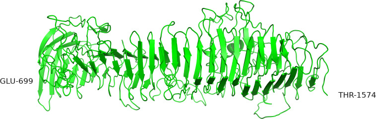
The cell envelope proteinase (CEP) of Lactococcus lactis is a large extracellular protease covalently linked to the peptidoglycan of the cell wall. Strains of L. lactis are typically auxotrophic for several amino acids and in order to grow to high cell densities in milk they need an extracellular protease. The structure of the entire CEP enzyme is difficult to determine experimentally due to the large size and due to the attachment to the cell surface. We here describe the use of a combination of structure prediction tools to create a structural model for the entire CEP enzyme of Lactococcus lactis. The model has implications for how the bacterium interacts with casein micelles during growth in milk, and it has implications regarding the energetics of the proteolytic system. Our model for the CEP indicates that the catalytic triad is activated through a structural change caused by interaction with the substrate. The CEP of L. lactis might become a useful model for the mode of action for enzymes belonging to the large class of S8 proteinases with a PA (protease associated) domain and a downstream fibronectin like domain.

Keywords: S8 proteinase; casein micelle; cell envelope associated peptidases; lactic acid bacteria (LAB); protein structure prediction; proteolytic system; substrate specificity; subtilisin.


Conflict of interest statement

The authors declare that the research was conducted in the absence of any commercial or financial relationships that could be construed as a potential conflict of interest.

Figures

FIGURE 1
FIGURE 1
Contact map for L. lactis MS22337 PrtP generated by RaptorX by superimposing 799 aa long segments of the PrtP sequence starting from the first aa of the mature protein (D186). As the map covers only the region around the diagonal, the composite map has been tilted by 45°. The four segments are 1–799, 301–1099, 501–1299, and 926–1725 (in this figure, positions are relative to D186).
FIGURE 2
FIGURE 2
Predicted structures of the MS22337 PrtP proteinase for the domain spanning residue nr 186–698. The superimposed structures are shown for iTAS_1 in green, iTAS_3 in cyan, iTAS_4 in yellow, Phyre2 in salmon, and Swiss in gray. The protein backbones are shown as cartoons with the catalytic triad shown as stick models with D in red, H in magenta, and S in blue.
FIGURE 3
FIGURE 3
The catalytic triad of MS22337 PrtP in the six structural models predicted based on the amino acid sequence. Aspartic acid, D215, is shown in red; Histidine, H279, is shown in magenta; and Serine, S618, is shown in blue. The protein backbone of the α carbons are shown as tubes with different color: iTAS_1 in green, iTAS_2 in light blue, iTAS_3 in dark gray, iTAS_4 in yellow, Phyre2 in amber, and Swiss in light gray. All six structures have the aspartic acid (215) at the same position and they have the serine (618) at coinciding positions in a distance of 7 Å from the aspartic acid. The histidine (279) of four models (iTAS_2, iTAS_3, iTAS_4, and Swiss) are located a the same position in a distance of 6 Å from the two other amino acids of the triad, whereas the histidines of iTAS_1 and Phyre2 are located far from the active site with distances of 18 and 15 Å respectively to the S618.
FIGURE 4
FIGURE 4
The alternative structures for the segment containing the histidine (279) of the catalytic triad. Four of the six structures have identical structures and are all shown in cyan whereas the two deviating structures iTAS_1 and Phyre2 are shown in green and salmon respectively. His 279s are shown as stick models in all structures.
FIGURE 5
FIGURE 5
Substrate position in the catalytic cleft of PrtP. (A) The iTAS4 model. The pro-peptide (shown as cartoon in red) is positioned in the catalytic cleft on iTAS_4 (shown as green surface. The catalytic triad is shown in yellow. (B) The obstructions in path of substrate posed by the iTAS_3 model is shown as magenta surfaces in the same model as (A). (C) The underlying change in structure allowing the substrate access to the catalytic cleft. Residue 320 is obstructing to one side and residue 429 is obstructing at the other site. The segments from 300–340 and 422–445 of iTAS_3 and iTAS_4 are shown as cartoons in magenta and green respectively inside the same surface as (B) (now transparent). (D) Possible substrate interaction in the area of fibronectin like structures. The surface of the iTAS_3 structure is shown in magenta. The position of substrate is shown in red and the catalytic triad is shown in yellow. The same obstacles for the substrate as seen in (C) is also obvious in this figure. The area in blue is a region within the fibronectin like domains spanning residue 930–950. This area might interact with larger substrates like entire caseins or long peptides.
FIGURE 6
FIGURE 6
Possible fibronectin like structure of the B domain established as a superposition of four overlapping structures build by Swiss model using a monomer of 4p99 as template. Only the six last units of the nine (based on Figure 1) fibronectin like structures could be fitted to the 4p99 template and only the five last gave a complete fit to this structure. The four overlapping structures are colored in yellow, green, cyan, and magenta.
FIGURE 7
FIGURE 7
Alternative structure of the AB domain modeled by iTASSER. In this structure the β-sheets are organized in parallel orientations.
FIGURE 8
FIGURE 8
The H domain of Lactococcus lactis PrtP modeled by RaptorX.
FIGURE 9
FIGURE 9
Model for the surface bound PrtP enzyme (shown in green) hydrolyzing caseins within the casein micelle. PrtP is covalently attached to the surface of the bacterium through a C-terminal threonine joined to peptidoglycan. The height of the enzyme is approximately 40 nm with the N-terminal proteinase domain at the top of molecule. The casein micelle (shown in red) has a diameter of 100 nm and is presented as the “Holt model” (Holt, 2016). We propose that the bacterium attach to the surface of the micelle and thereby position the proteinase domain in an environment of rich in casein substrates. The oligopeptide transporter proteins OppA, OppB, OppC, OppD, and OppF are shown as cartoons inspired by Doeven et al. (2005).
FIGURE 10
FIGURE 10
Position of the dispensable loop of Bruinenberg et al. (1994b). The structure of PrtP iTAS_4 model is shown as surface in green. Substrate positioned in the catalytic cleft is shown as red cartoon. The catalytic triad is shown as yellow on the surface of the enzyme. (A) The loop from 423 to 573 is shown in magenta, on (B) the entire loop has been removed. Residues 422 and 574 are quite close in the model (10 Å distance).

Similar articles

Cited by

References

    1. Abdelgadir W., Nielsen D. S., Hamad S., Jakobsen M. (2008). A traditional Sudanese fermented camel’s milk product, gariss, as a habitat of Streptococcus infantarius subsp. infantarius. Int. J. Food Microbiol. 127 215–219. 10.1016/j.ijfoodmicro.2008.07.008 - DOI - PubMed
    1. Bachmann H., Starrenburg M. J. C., Molenaar D., Kleerebezem M., van Hylckama Vlieg J. E. T. (2012). Microbial domestication signatures of Lactococcus lactis can be reproduced by experimental evolution. Genome Res. 22 115–124. 10.1101/gr.121285.111 - DOI - PMC - PubMed
    1. Balder R., Lipski S., Lazarus J. J., Grose W., Wooten R. M., Hogan R. J., et al. (2010). Identification of Burkholderia mallei and Burkholderia pseudomallei adhesins for human respiratory epithelial cells. BMC Microbiol. 10:250. 10.1186/1471-2180-10-250 - DOI - PMC - PubMed
    1. Bergman C., Kashiwaya Y., Veech R. L. (2010). The effect of pH and free Mg2+ on ATP linked enzymes and the calculation of Gibbs free energy of ATP hydrolysis. J. Phys. Chem. B 114 16137–16146. 10.1021/jp105723r - DOI - PubMed
    1. Berhe T., Ipsen R., Seifu E., Kurtu M. Y., Eshetu M., Hansen E. B. (2018). Comparison of the acidification activities of commercial starter cultures in camel and bovine milk. LWT 89 123–127. 10.1016/j.lwt.2017.10.041 - DOI