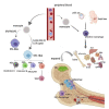
Communications Between Bone Marrow Macrophages and Bone Cells in Bone Remodeling
- PMID: 33415105
- PMCID: PMC7783313
- DOI: 10.3389/fcell.2020.598263
Communications Between Bone Marrow Macrophages and Bone Cells in Bone Remodeling
Abstract
The mammalian skeleton is a metabolically active organ that continuously undergoes bone remodeling, a process of tightly coupled bone resorption and formation throughout life. Recent studies have expanded our knowledge about the interactions between cells within bone marrow in bone remodeling. Macrophages resident in bone (BMMs) can regulate bone metabolism via secreting numbers of cytokines and exosomes. This review summarizes the current understanding of factors, exosomes, and hormones that involved in the communications between BMMs and other bone cells including mensenchymal stem cells, osteoblasts, osteocytes, and so on. We also discuss the role of BMMs and potential therapeutic approaches targeting BMMs in bone remodeling related diseases such as osteoporosis, osteoarthritis, rheumatoid arthritis, and osteosarcoma.
Keywords: bone diseases; bone remodeling; intracellular communication; macrophage-derived exosome; macrophages.
Copyright © 2020 Chen, Jiao, Liu, Huang, He, He, Hou, Yang, Luo and Li.
Conflict of interest statement
The authors declare that the research was conducted in the absence of any commercial or financial relationships that could be construed as a potential conflict of interest.
Figures


Similar articles
-
Reversal of Osteoporotic Activity by Endothelial Cell-Secreted Bone Targeting and Biocompatible Exosomes.Nano Lett. 2019 May 8;19(5):3040-3048. doi: 10.1021/acs.nanolett.9b00287. Epub 2019 Apr 23. Nano Lett. 2019. PMID: 30968694
-
Exosomes: mediators of bone diseases, protection, and therapeutics potential.Oncoscience. 2018 Jun 23;5(5-6):181-195. doi: 10.18632/oncoscience.421. eCollection 2018 May. Oncoscience. 2018. PMID: 30035185 Free PMC article. Review.
-
Bone remodeling.Ann N Y Acad Sci. 2006 Dec;1092:385-96. doi: 10.1196/annals.1365.035. Ann N Y Acad Sci. 2006. PMID: 17308163 Review.
-
Bone remodeling in the context of cellular and systemic regulation: the role of osteocytes and the nervous system.J Mol Endocrinol. 2015 Oct;55(2):R23-36. doi: 10.1530/JME-15-0067. Epub 2015 Aug 25. J Mol Endocrinol. 2015. PMID: 26307562 Review.
-
Reactivity of rat bone marrow-derived macrophages to neurotransmitter stimulation in the context of collagen II-induced arthritis.Arthritis Res Ther. 2015 Jun 24;17(1):169. doi: 10.1186/s13075-015-0684-4. Arthritis Res Ther. 2015. PMID: 26104678 Free PMC article.
Cited by
-
Inflammasomes and their roles in arthritic disease pathogenesis.Front Mol Biosci. 2022 Oct 28;9:1027917. doi: 10.3389/fmolb.2022.1027917. eCollection 2022. Front Mol Biosci. 2022. PMID: 36387275 Free PMC article. Review.
-
Caspase-resistant ROCK1 expression prolongs survival of Eµ-Myc B cell lymphoma mice.Dis Model Mech. 2024 May 1;17(5):dmm050631. doi: 10.1242/dmm.050631. Epub 2024 May 21. Dis Model Mech. 2024. PMID: 38616733 Free PMC article.
-
From Synthesis to Clinical Trial: Novel Bioinductive Calcium Deficient HA/β-TCP Bone Grafting Nanomaterial.Nanomaterials (Basel). 2023 Jun 17;13(12):1876. doi: 10.3390/nano13121876. Nanomaterials (Basel). 2023. PMID: 37368306 Free PMC article.
-
Roles of Altered Macrophages and Cytokines: Implications for Pathological Mechanisms of Postmenopausal Osteoporosis, Rheumatoid Arthritis, and Alzheimer's Disease.Front Endocrinol (Lausanne). 2022 Jun 10;13:876269. doi: 10.3389/fendo.2022.876269. eCollection 2022. Front Endocrinol (Lausanne). 2022. PMID: 35757427 Free PMC article. Review.
-
Effects of Estrogens on Osteoimmunology: A Role in Bone Metastasis.Front Immunol. 2022 May 23;13:899104. doi: 10.3389/fimmu.2022.899104. eCollection 2022. Front Immunol. 2022. PMID: 35677054 Free PMC article. Review.
References
Publication types
LinkOut - more resources
Full Text Sources

