. 2021 Feb;18(1):143-154.
doi: 10.1007/s13770-020-00312-1. Epub 2021 Jan 7.

Human Dental Pulp Stem Cells (DPSCs) Therapy in Rescuing Photoreceptors and Establishing a Sodium Iodate-Induced Retinal Degeneration Rat Model

Affiliations

Human Dental Pulp Stem Cells (DPSCs) Therapy in Rescuing Photoreceptors and Establishing a Sodium Iodate-Induced Retinal Degeneration Rat Model

Chenshen Lam et al. Tissue Eng Regen Med. 2021 Feb.

Abstract

Background: Different methods have been used to inject stem cells into the eye for research. We previously explored the intravitreal route. Here, we investigate the efficacy of intravenous and subretinal-transplanted human dental pulp stem cells (DPSCs) in rescuing the photoreceptors of a sodium iodate-induced retinal degeneration model.

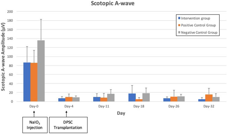
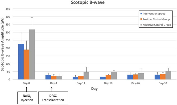
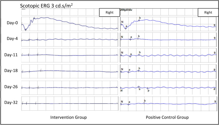
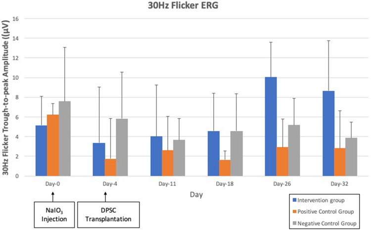
Methods: Three groups of Sprague Dawley rats were used: intervention, vehicle group and negative control groups (n = 6 in each). Intravenous injection of 60 mg/kg sodium iodate (day 0) induced retinal degeneration. On day 4 post-injection of sodium iodate, the rats in the intervention group received intravenous DPSC and subretinal DPSC in the right eye; rats in the vehicle group received subretinal Hank's balance salt solution and intravenous normal saline; while negative control group received nothing. Electroretinogram (ERG) was performed to assess the retinal function at day 0 (baseline), day 4, day 11, day 18, day 26, and day 32. By the end of the study at day 32, the rats were euthanized, and both their enucleated eyes were sent for histology.

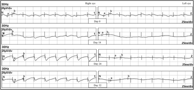
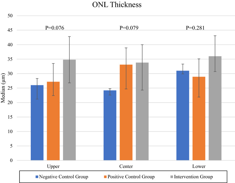
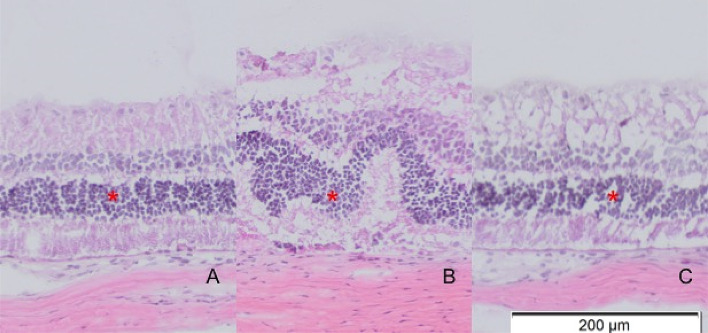
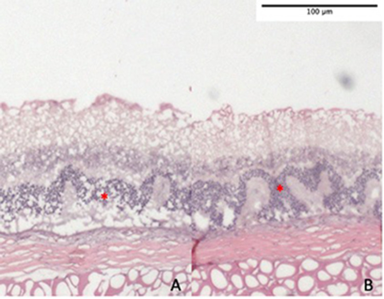
Results: No significant difference in maximal ERG a-wave (p = 0.107) and b-wave, (p = 0.153) amplitude was seen amongst the experimental groups. However, photopic 30 Hz flicker amplitude of the study eye showed significant differences in the 3 groups (p = 0.032). Within the intervention group, there was an improvement in 30 Hz flicker ERG response of all 6 treated right eyes, which was injected with subretinal DPSC; while the 30 Hz flicker ERG of the non-treated left eyes remained flat. Histology showed improved outer nuclear layer thickness in intervention group; however, findings were not significant compared to the negative and vehicle groups.

Conclusion: Combination of subretinal and intravenous injection of DPSCs may have potential to rescue cone function from a NaIO3-induced retinal injury model.

Keywords: Degenerated retina; Dental pulp; Electroretinography; Mesenchymal stem cell; Sodium iodate; Sprague-Dawley rats.


Conflict of interest statement

The authors declare no conflict of interest.

Figures

Fig. 1
Fig. 1
Scotopic 3 cd.s/m2 a-wave amplitude of intervention group, vehicle group group and negative control group. There was reduction of the amplitude from day 4 to day 32 after NaIO3 injection in all 3 groups. No significant difference is seen in a-wave amplitude amongst the experimental groups at all points in time (p = 0.107)
Fig. 2
Fig. 2
Scotopic 3 cd.s/m2 b-wave amplitude in the intervention group, vehicle group group and negative control groups. There was reduction of the amplitude from day 4 to day 32 after NaIO3 injection in all 3 groups with no significant differences (p = 0.153)
Fig. 3
Fig. 3
Scotopic 3 cd.s/m2 ERG strip of both the intervention group and negative control group throughout the study, showing similar responses at all study points
Fig. 4
Fig. 4
30 Hz Flicker ERG of right eye and left eye of SD rat in intervention group. There were regular flicker waves appearing from day 18 onwards to day 32 over right eye only which is the eye injected with subretinal DPSC. However, the waveform was not similar to those at baseline on day 0. The left eye, on the other hand, remains flat throughout the study
Fig. 5
Fig. 5
30 Hz flicker ERG trough-to-peak amplitude of intervention group, vehicle group and negative control group with significant difference (p = 0.032)
Fig. 6
Fig. 6
ONL thickness of upper peripheral retina, central retina, and lower peripheral retina of both intervention group and negative control group
Fig. 7
Fig. 7
AC Histology of intervention group showing some preservation of ONL thickness in upper peripheral retina (A) and lower peripheral retina (C) in comparison to central retina (B) with less corrugation and disruption of ONL indicated by red asterisks
Fig. 8
Fig. 8
A, B Histology of the central retina from negative controls (A) and vehicle groups (B) showing corrugation and thinning of ONL due to NaIO3 toxicity. ONL is indicated by red asterisks

Similar articles

Cited by

References

    1. Leow SN, Luu CD, Hairul Nizam MH, Mok PL, Ruhaslizan R, Wong HS, et al. Safety and efficacy of human Wharton’s Jelly-derived mesenchymal stem cells therapy for retinal degeneration. PLoS One. 2015;10:e0128973. doi: 10.1371/journal.pone.0128973. - DOI - PMC - PubMed
    1. Jiang Y, Zhang Y, Zhang L, Wang M, Zhang X, Li X. Therapeutic effect of bone marrow mesenchymal stem cells on laser-induced retinal injury in mice. Int J Mol Sci. 2014;15:9372–9385. doi: 10.3390/ijms15069372. - DOI - PMC - PubMed
    1. Machalinska A, Roginska D, Pius-Sadowska E, et al. Neuroprotective and antiapoptotic activity of lineage-negative bone marrow cells after intravitreal injection in a mouse model of acute retinal injury. Stem Cells Int. 2015;2015:620364. doi: 10.1155/2015/620364. - DOI - PMC - PubMed
    1. Zhang Y, Wang W. Effects of bone marrow mesenchymal stem cell transplantation on light-damaged retina. Invest Ophthalmol Vis Sci. 2010;51:3742–3748. doi: 10.1167/iovs.08-3314. - DOI - PubMed
    1. Haddad-Mashadrizeh A, Bahrami AR, Matin MM, Edalatmanesh MA, Zomorodipour A, Gardaneh M, et al. Human adipose-derived mesenchymal stem cells can survive and integrate into the adult rat eye following xenotransplantation. Xenotransplantation. 2013;20:165–176. - PubMed

Publication types

LinkOut - more resources