. 2021 May;238(5):1082-1105.
doi: 10.1111/joa.13362. Epub 2021 Jan 7.

The morphology and evolution of chondrichthyan cranial muscles: A digital dissection of the elephantfish Callorhinchus milii and the catshark Scyliorhinus canicula

Affiliations

The morphology and evolution of chondrichthyan cranial muscles: A digital dissection of the elephantfish Callorhinchus milii and the catshark Scyliorhinus canicula

Richard P Dearden et al. J Anat. 2021 May.

Erratum in

  • Corrigendum.
    [No authors listed] [No authors listed] J Anat. 2021 Oct;239(4):972. doi: 10.1111/joa.13498. Epub 2021 Jul 26. J Anat. 2021. PMID: 34312847 Free PMC article. No abstract available.

Abstract

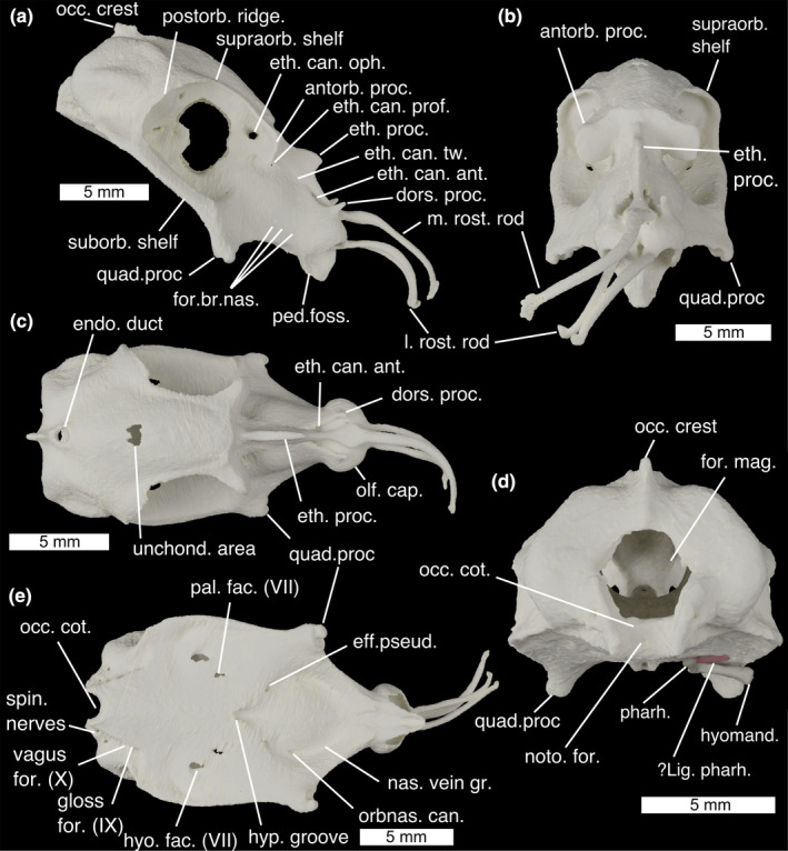
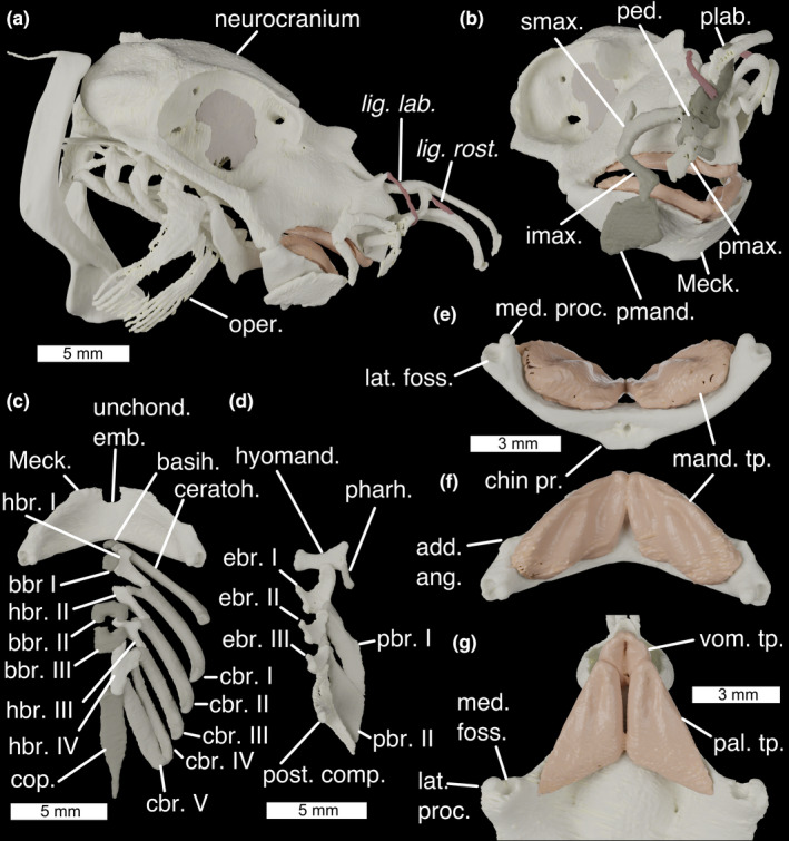
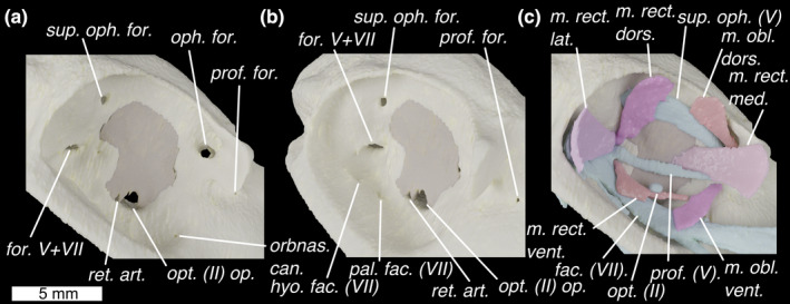
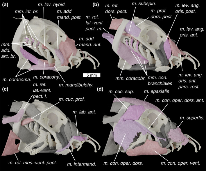
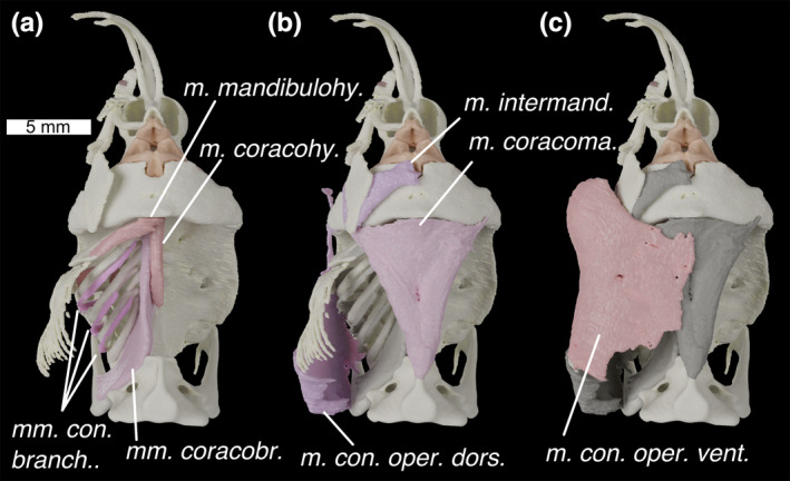
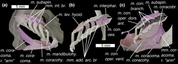
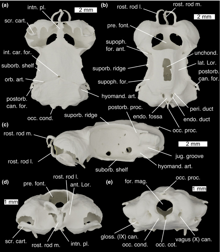
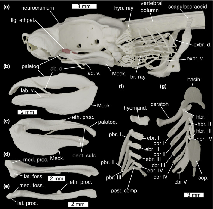
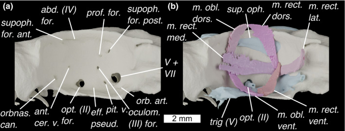
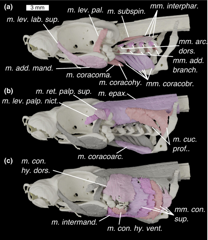
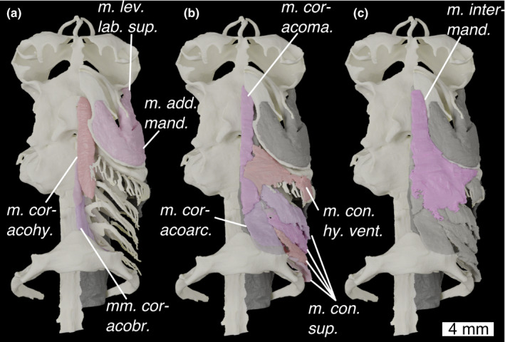
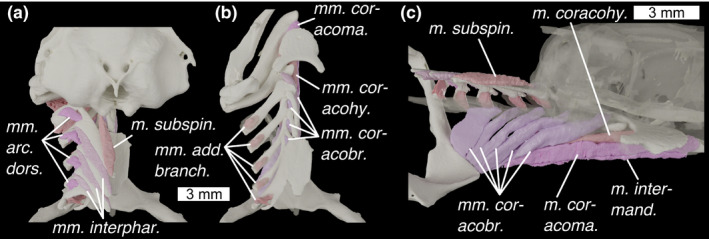
The anatomy of sharks, rays, and chimaeras (chondrichthyans) is crucial to understanding the evolution of the cranial system in vertebrates due to their position as the sister group to bony fishes (osteichthyans). Strikingly different arrangements of the head in the two constituent chondrichthyan groups-holocephalans and elasmobranchs-have played a pivotal role in the formation of evolutionary hypotheses targeting major cranial structures such as the jaws and pharynx. However, despite the advent of digital dissections as a means of easily visualizing and sharing the results of anatomical studies in three dimensions, information on the musculoskeletal systems of the chondrichthyan head remains largely limited to traditional accounts, many of which are at least a century old. Here, we use synchrotron tomographic data to carry out a digital dissection of a holocephalan and an elasmobranch widely used as model species: the elephantfish, Callorhinchus milii, and the small-spotted catshark, Scyliorhinus canicula. We describe and figure the skeletal anatomy of the head, labial, mandibular, hyoid, and branchial cartilages in both taxa as well as the muscles of the head and pharynx. In Callorhinchus, we make several new observations regarding the branchial musculature, revealing several previously unreported or ambiguously characterized muscles, likely homologous to their counterparts in the elasmobranch pharynx. We also identify a previously unreported structure linking the pharyngohyal of Callorhinchus to the neurocranium. Finally, we review what is known about the evolution of chondrichthyan cranial muscles from their fossil record and discuss the implications for muscle homology and evolution, broadly concluding that the holocephalan pharynx is likely derived from a more elasmobranch-like form which is plesiomorphic for the chondrichthyan crown group. This dataset has great potential as a resource, particularly for researchers using these model species for zoological research, functional morphologists requiring models of musculature and skeletons, as well as for palaeontologists seeking comparative models for extinct taxa.

Keywords: Callorhinchus milii; Scyliorhinus canicula; cranial muscles; digital dissection; elasmobranch; holocephalan.


Figures

FIGURE 1
FIGURE 1
The neurocranium of Callorhinchus milii in (a) lateral, (b) anterior, (c) dorsal, (d) posterior and (e) ventral views. ?lig. pharh., ?ligamentum pharyngohyoideus; antorb. proc., antorbital process; dors. proc. dorsal process; eff. pseud., foramen for the efferent pseudobranchial artery; endo. duct, foramen for endolymphatic duct; eth. can. Ant, anterior ethmoid canal foramina eth. can. oph, entry into ethmoid canal for superficial ophthalmic complex; eth. can. prof, entry into ethmoid canal for profundus nerve; eth. can. tw., exit from ethmoid canal for twigs of the superficial ophthalmic complex/profundus; eth. proc., ethmoid process; for. br. nas., foramina for branches of the nasal vein; for. mag., foramen magnum; gloss for. (IX), foramen for glossopharyngeal (IX) nerve; hyo. fac. (VII), foramen for hyomandibular branch of the facial (VII) nerve; hyomand., hyomandibula; hyp. groove., hypophyseal groove; l. rost. rod, lateral rostral rod; m. rost. rod, median rostral rod; nas. vein gr., groove for the nasal vein; noto. for., notochord foramen; occ. cot., occipital cotylus; occ. crest, occipital crest; olf. cap., olfactory capsules; orbnas. can., foramen for the orbitonasal canal; pal. fac. (VII), foramen for palatine branch of the facial (VII) nerve; ped. foss., fossa for pedicular cartilage; pharh, pharyngohyal; postorb. ridge, postorbital ridge; quad. proc., quadrate process; spin. nerves, foramina for anterior spinal nerves; suborb. shelf, suborbital shelf; supraorb. shelf, supraorbital shelf; unchond. area, incompletely chondrified area; vagus for. (X), foramen for vagus (X) nerve
FIGURE 2
FIGURE 2
The cranial skeleton of Callorhinchus milii (a) complete skeleton in lateral view, (b) antero‐lateral view showing labial cartilages, (c) ventral pharyngeal skeleton in dorsal view, (d) dorsal pharyngeal skeleton in ventral view, lower jaw in ventral (e) and dorsal (f) view, and (g) palate in ventral view. Colours: cream, cartilage; beige, sphenoptic membrane; red, ligaments. add. ang., adductor mandibulae angle; basih., basihyal; bbr., basibranchials; cbr., ceratobranchial; ceratoh, ceratohyal; chin pr., chin process; cop., basibranchial copula; ebr, epibranchial; hbr., hypobranchial; hyomand., hyomandibula; imax., inferior maxillary cartilage; lat. foss., lateral fossa of Meckel's cartilage; lat. proc., lateral process of quadrate; lig. lab., ligamentum labialis; lig. rost., ligamentum rostralis; mand. tp., mandibular toothplates; Meck., Meckel's cartilage; med. foss, medial fossa of quadrate; med. proc., medial process of Meckel's cartilage; oper., opercular cartilage; pal. tp. palatine toothplates; pbr., pharyngobranchial; ped. pedicular cartilage; pharh, pharyngohyal; plab., prelabial cartilage; pmand. premandibular cartilage; pmax., premaxillary cartilage; post. comp., posterior epibranchial/pharyngobranchial complex; smax., superior maxillary cartilage; unchond. emb., unchondrified embayment; vom. tp., vomerine toothplates
FIGURE 3
FIGURE 3
The orbit of Callorhinchus milii shown in lateral view with skeleton in (a) lateral and (b) antero‐lateral view and (c) lateral view with external optic muscles and cranial nerves. Colours as in Figure 3 with light‐blue for cranial nerves. fac. (VII), facial (VII) nerve; for., foramen for superficial ophthalmic complex's entry into orbit; for. V+VII, foramen for entry of trigeminal (V) and facial (VII) nerves into the orbit; hyo. fac. (VII), foramen for hyomandibular branch of the facial (VII) nerve; m. obl. dors., m. obliquus dorsalis; m. obl. vent., m. obliquus ventralis; m. rect. dors., m. rectus dorsalis; m. rect. lat., m. rectus lateralis; m. rect. med., m. rectus medialis; m. rect. vent., m. rectus ventralis; oph. for. ophthalmic foramen; opt. (II) op., optic nerve opening; opt. (II), optic nerve; orbnas. can., foramen for the orbitonasal canal; pal. fac. (VII), foramen for palatine branch of the facial (VII) nerve; prof. (V), profundus (V) nerve; prof. for., foramen for profundus; ret. art., retinal artery opening; sup. oph. (V), superficial ophthalmic complex (V+anterodorsal lateral line nerves); sup. oph
FIGURE 4
FIGURE 4
Lateral view of the head of Callorhinchus milii (a–d) showing progressively more shallow muscles with previous layer/s of muscles shown in grey. Colours: cream, cartilage; beige, sphenoptic membrane; red, ligaments; pinks, muscles; greys, deeper muscles. m. add. mand. ant., m. adductor mandibulae anterior; m. add. mand. post., m. adductor mandibulae posterior; m. con. oper. dors., m. constrictor opercula dorsalis; m. con. oper. dors. ant., m. constrictor operculi dorsalis anterior; m. con. oper. vent., m. constrictor operculi ventralis; m. coracohy., mm. coracohyoideus; m. coracoma., m. coracomandibularis (coloured in two shades to show division); m. cuc. prof., m. cucullaris profundus; m. cuc. sup., m. cucullaris superficialis; m. intermand., m. intermandibularis; m. lab. ant., m. labialis anterior; m. lev. ang. oris ant. pars. rost., m. levator anguli oris anterior pars rostralis; m. lev. ang. oris ant., m. levator anguli oris anterior; m. lev. ang. oris post., levator anguli oris posterior; m. lev. hyoid., m. levator hyoideus; m. mandibulohy., m. mandibulohyoideus; m. prot. dors. pect., m. protractor dorsalis pectoralis; m. ret. dors. pect., m. retractor dorsalis pectoralis; m. ret. lat.‐vent. pect. l, m. retractor latero‐ventralis pectoralis lateral; m. ret. lat.‐vent. pect. m, m. retractor latero‐ventralis pectoralis medial; m. ret. mes.‐vent. pect., m. retractor mesio‐ventralis pectoralis; m. subspin, m. subspinalis; m. superfic., m. superficialis; mm. add. arc. br., mm. adductors arcuum branchiales; mm. con. branchiales, mm. constrictors branchiales; mm. coracobr., mm. coracobranchiales; mm. int. br., m. interarcuales branchiales
FIGURE 5
FIGURE 5
Ventral view of the head of Callorhinchus milii with (a) deepest muscles, (b) deeper muscles and (c) shallow muscles overlain. Colours and abbreviations as in Figure 4
FIGURE 6
FIGURE 6
Branchial skeleton of Callorhinchus milii with (a) in postero‐lateral view with semi‐transparent neurocranium and scapulocoracoid, (b) in medial view and (c) in antero‐medial view with a semi‐transparent mandible. Colours and abbreviations as in Figure 3. Additional abbreviations: m. con. oes., m. constrictor oesophagi; m. coracoma. r. arm, right “arm” of m. coracomandibularis (left not pictured); m. interphar; m. interpharyngobranchialis. Otherwise as for Figure 4
FIGURE 7
FIGURE 7
The neurocranium of Scyliorhinus canicula in (a) ventral, (b) dorsal, (c) lateral, (d) anterior and (e) posterior views. ant. Lor., anterior depression for ampullae of Lorenzini; endo. duct, endolymphatic duct; endo. fossa, endolymphatic fossa; for. mag., foramen magnum; gloss. (IX) can., glossopharyngeal (IX) nerve canal exit; hyomand. art, hyomandibular articulation surface.; int. car. for., foramen for the internal carotids; intn. pl., internasal plate; jug. groove, jugular groove; lat. Lor., lateral furrows for ampullae of Lorenzini; occ. cond., occipital condyle; occ. cot., occipital cotylus; occ. proc., occipital processes; orb. art., foramen for the orbital artery; peri. duct, perilymphatic duct; postorb. can. for., foramen for postorbital sensory canal; postorb. can. for., foramen for postorbital sensory canal; postorb. proc., postorbital process; pre. font., precerebral fontanelle; rost. rod l, lateral rostral rod; rost. rod m., median rostral rod; scr. cart., scrolled cartilage; suborb. shelf; supoph. for. ant., anterior foramen for the superophthalmic complex; supoph. for. foramina for twigs of the superophthalmic complex; supoph. for., foramina for the superophthalmic complex; suporb. ridge, supraorbital ridge; unchond., incompletely chondrified area; vagus (X) can., vagus (X) nerve canal exit
FIGURE 8
FIGURE 8
Cranial skeleton of Scyliorhinus canicula with (a) whole cranial skeleton in lateral view, mandibles in (b) lateral and (c) medial view, (d) left Meckel's cartilage in dorsal view, (e) left palatoquadrate in ventral view, (f) dorsal gill skeleton in ventral view and (g) ventral gill skeleton in dorsal view. basih., basihyal; br. ray, branchial rays; cbr., ceratobranchial; ceratoh, ceratohyal; cop., basibranchial copula; dent. sulc., dental sulcus; ebr, epibranchial; eth. proc., ethmoid process; exbr. d., dorsal extrabranchial cartilages; exbr. v., ventral extrabranchial cartilages; hbr., hypobranchial; hyo. ray, hyoid rays; hyomand., hyomandibula; lab. d., dorsal labial cartilage; lab. v., ventral labial cartilage; lat. foss., lateral fossa of Meckel's cartilage; lat. proc., lateral process of palatoquadrate; Meck., Meckel's cartilage; med. foss, medial fossa of palatoquadrate; med. proc., medial process of Meckel's cartilage; palatoq., palatoquadrate; pbr., pharyngobranchial; post. comp., posterior epibranchial/pharyngobranchial complex
FIGURE 9
FIGURE 9
The orbit of Scyliorhinus canicula shown in lateral view with (a) foramina shown, and (b) external optic muscles and cranial nerves. Colours as in Figure 9 with light‐blue for cranial nerves. abd (IV) for., foramen for the abducens (IV) nerve; ant. cer. v., foramen for the anterior cerebral vein; eff. pseud, foramen for the efferent pseudobranchial artery; m. obl. dors., m. obliquus dorsalis; m. obl. vent., m. obliquus ventralis; m. rect. dors., m. rectus dorsalis; m. rect. lat., m. rectus lateralis; m. rect. med., m. rectus medialis; m. rect. vent., m. rectus ventralis; oculom. (III) for., foramen for the oculomotor (III) nerve; opt (II), optic nerve; opt. (II) for., foramen for the optic (II) nerve; orb. art., foramen for the orbital artery; orbnas. can., foramen for the orbitonasal canal; pit. v., foramen for pituitary vein; prof. for., foramen for profundus; sup. oph., superficial ophthalmic complex (V); supoph. for. ant., anterior foramen for the superophthalmic complex; supoph. for. post., posterior foramen for the superophthalmic complex; trig. (V), trigeminal nerve; V+VII, foramen for entry of trigeminal (V) and facial (VII) nerves into the orbit
FIGURE 10
FIGURE 10
Lateral view of the head of Scyliorhinus canicula (a–c) showing progressively more shallow muscles with previous layer/s of muscles shown in grey. Colours: cream, cartilage; beige, pinks, muscles; greys, deeper muscles. m. add. mand., m. adductor mandibulae; m. con. hy. dors., m. constrictor hyoideus dorsalis; m. con. hy. vent., m. constrictor hyoideus ventralis; m. coracoarc., m. coracoarcualis; m. coracohy., m. coracohyoideus; m. coracoma., m. coracomandibularis; m. cuc. prof., m. cucullaris profundus; m. epax., m. epaxialis; m. intermand., m. intermandibularis; m. lev. lab. sup., m. levator labii superioris; m. lev. pal., m. levator palatoquadrate; m. lev. palp. nict., m. levator palpebrae nictitantis; m. ret. palp. sup., m. retractor palpebrae superioris; m. subspin., m. subspinalis; mm. add. branch., m. adductors branchiales; mm. arc. dors., mm. arcuales branchiales; mm. con. sup., mm. constrictors superficiales; mm. coracobr., mm. coracobranchiales; mm. interphar., mm. interpharyngobranchiales
FIGURE 11
FIGURE 11
Ventral view of the head of Scyliorhinus canicula with (a) deepest muscles, (b) deeper muscles and (c) shallow muscles overlain. Colours and abbreviations as in Figure 10
FIGURE 12
FIGURE 12
Branchial skeleton of Scyliorhinus canicula (a) in dorsal view, with neurocranium (b) in dorsal view with neurocranium removed and dorsal branchial skeleton semi‐transparent, and (c) in medial view with nerocranium and central gill‐skeleton semi‐transparent. Colours and abbreviations as in Figure 10

Similar articles

Cited by

References

    1. Allis, E.P. (1897) The cranial muscles of Amia . Journal of Morphology, XII, 489–772.
    1. Allis, E.P. (1917) The homologies of the muscles related to the visceral arches of the gnathostome fishes. Quarterly Journal of Microscopical Science, 62, 303–406.
    1. Anderson, P.S.L. (2008) Cranial muscle homology across modern gnathostomes. Biological Journal of the Linnean Society, 94, 195–216.
    1. Brazeau, M.D. & de Winter, V. (2015) The hyoid arch and braincase anatomy of Acanthodes support chondrichthyan affinity of ‘acanthodians’. Proceedings of the Royal Society B: Biological Sciences, 282, 20152210. - PMC - PubMed
    1. Brocklehurst, R. , Porro, L. , Herrel, A. , Adriaens, D. & Rayfield, E.J. (2019) A digital dissection of two teleost fishes: comparative functional anatomy of the cranial musculoskeletal system in pike (Esox lucius) and eel (Anguilla anguilla). Journal of Anatomy, 235, 189–204. - PMC - PubMed

Publication types

MeSH terms

LinkOut - more resources