Randomized Controlled Trial
. 2021 Mar;121(3):941-955.
doi: 10.1007/s00421-020-04582-4. Epub 2021 Jan 8.

Insights into the combination of neuromuscular electrical stimulation and motor imagery in a training-based approach

Affiliations
Randomized Controlled Trial

Insights into the combination of neuromuscular electrical stimulation and motor imagery in a training-based approach

Amandine Bouguetoch et al. Eur J Appl Physiol. 2021 Mar.

Abstract

Introduction: Training stimuli that partially activate the neuromuscular system, such as motor imagery (MI) or neuromuscular electrical stimulation (NMES), have been previously shown as efficient tools to induce strength gains. Here the efficacy of MI, NMES or NMES + MI trainings has been compared.

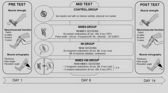
Methods: Thirty-seven participants were enrolled in a training program of ten sessions in 2 weeks targeting plantar flexor muscles, distributed in four groups: MI, NMES, NMES + MI and control. Each group underwent forty contractions in each session, NMES + MI group doing 20 contractions of each modality. Before and after, the neuromuscular function was tested through the recording of maximal voluntary contraction (MVC), but also electrophysiological and mechanical responses associated with electrical nerve stimulation. Muscle architecture was assessed by ultrasonography.

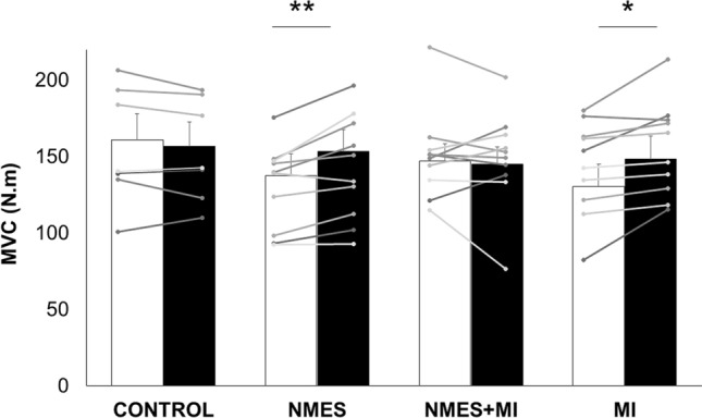
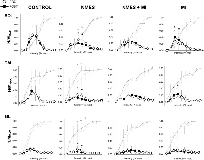
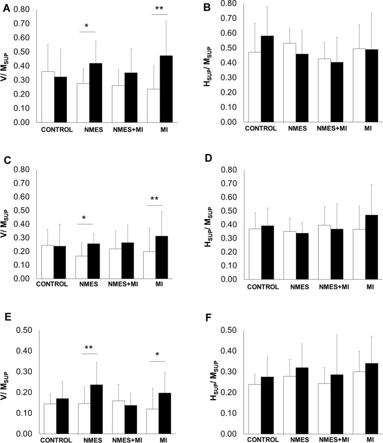
Results: MVC increased by 11.3 ± 3.5% in NMES group, by 13.8 ± 5.6% in MI, while unchanged for NMES + MI and control. During MVC, a significant increase in V-wave without associated changes in superimposed H-reflex has been observed for NMES and MI, suggesting that neural adaptations occurred at supraspinal level. Rest spinal excitability was increased in the MI group while decreased in the NMES group. No change in muscle architecture (pennation angle, fascicle length) has been found in any group but muscular peak twitch and soleus maximal M-wave increased in the NMES group only.

Conclusion: Finally, MI and NMES seem to be efficient stimuli to improve strength, although both exhibited different and specific neural plasticity. On its side, NMES + MI combination did not provide the expected gains, suggesting that their effects are not simply cumulative, or even are competitive.

Keywords: Spinal excitability; Strength; Triceps surae; Ultrasonography; V-wave.


Conflict of interest statement

There is no conflict of interest or competing interests.

Figures

Fig. 1
Fig. 1
Overview of the experimental protocol
Fig. 2
Fig. 2
Plantar flexor muscles force characteristics of the four groups. Data are depicted as mean ± SD for the four groups: the control group (CONTROL) and the three trained groups: neuromuscular electrical stimulation only (NMES), combined NMES and motor imagery (NMES + MI) and MI only (MI). Maximal torque of the plantar flexor muscles recorded during maximal voluntary contraction (MVC) before (PRE, white bars) and after (POST, black bars) the training protocols. Individual data are depicted in gray dots and lines. *,**Significant PRE-POST difference at p < 0.05 and p < 0.01, respectively
Fig. 3
Fig. 3
Spinal excitability of the plantar flexor muscles in the four groups. Spinal excitability is expressed as the recruitment curves the H-reflexes and M-waves of the soleus (SOL), gastrocnemius medialis (GM) and gastrocnemius lateralis (GL) recorded for the four groups (CONTROL; NMES neuromuscular electrical stimulation only, MI motor imagery, NMES + MI combined training). Recruitment curves are built as follows: mean H-reflex responses are normalized by the corresponding maximal M-wave (MMAX) in ten portions of intensities (portion 1: from 0 to 10% of maximal intensity; portion 2: from 11 to 20% of maximal intensity; …; portion 10: from 91 to 100% of maximal intensity). Open circles are responses recorded at PRE, and full circles are responses recorded at POST. Associated M-waves to each H-reflex, also normalized by the corresponding maximal M-wave, are represented in light gray for PRE and dark gray for POST. *Significant PRE-to-POST response at p < 0.05 (dark: for H/MMAX and gray for MatH/MMAX)
Fig. 4
Fig. 4
Plantar flexors V-wave and superimposed H-reflex of the four groups. V/MSUP and HSUP/MSUP ratios are depicted for the four groups: the control group (CONTROL) and the three trained groups: neuromuscular electrical stimulation only (NMES), combined NMES and motor imagery (NMES + MI) and MI only (MI). Ratios before (PRE, white bars) and after (POST, black bars) are shown for the three tested muscles in a and b for soleus muscle, c and d for gastrocnemius medialis, and e and f for gastrocnemius lateralis. *,**Significant PRE-POST difference at p < 0.05 and p < 0.01, respectively

References

    1. Aagaard P. Training-induced changes in neural function. Exerc Sport Sci Rev. 2003;31:61–67. doi: 10.1097/00003677-200304000-00002. - DOI - PubMed
    1. Aagaard P, Simonsen EB, Andersen JL, et al. Neural adaptation to resistance training: changes in evoked V-wave and H-reflex responses. J Appl Physiol. 2002;92:2309–2318. doi: 10.1152/japplphysiol.01185.2001. - DOI - PubMed
    1. Bickel CS, Gregory CM, Dean JC. Motor unit recruitment during neuromuscular electrical stimulation: a critical appraisal. Eur J Appl Physiol. 2011;111:2399. doi: 10.1007/s00421-011-2128-4. - DOI - PubMed
    1. Carroll TJ, Selvanayagam VS, Riek S, Semmler JG. Neural adaptations to strength training: moving beyond transcranial magnetic stimulation and reflex studies. Acta Physiol. 2011;202:119–140. doi: 10.1111/j.1748-1716.2011.02271.x. - DOI - PubMed
    1. Cattagni T, Scaglioni G, Laroche D, et al. Ankle muscle strength discriminates fallers from non-fallers. Front Aging Neurosci. 2014 doi: 10.3389/fnagi.2014.00336. - DOI - PMC - PubMed

Publication types

LinkOut - more resources