. 2021 Feb;14(2):e009229.
doi: 10.1161/CIRCEP.120.009229. Epub 2021 Jan 8.

Circular Multielectrode Pulsed Field Ablation Catheter Lasso Pulsed Field Ablation: Lesion Characteristics, Durability, and Effect on Neighboring Structures

Affiliations

Circular Multielectrode Pulsed Field Ablation Catheter Lasso Pulsed Field Ablation: Lesion Characteristics, Durability, and Effect on Neighboring Structures

Hagai Yavin et al. Circ Arrhythm Electrophysiol. 2021 Feb.

Abstract

Background: Pulsed field ablation (PFA) is a nonthermal energy with potential safety advantages over radiofrequency ablation. This study investigated a novel PFA system-a circular multielectrode catheter (PFA lasso) and a multichannel generator designed to work with Carto 3 mapping system.

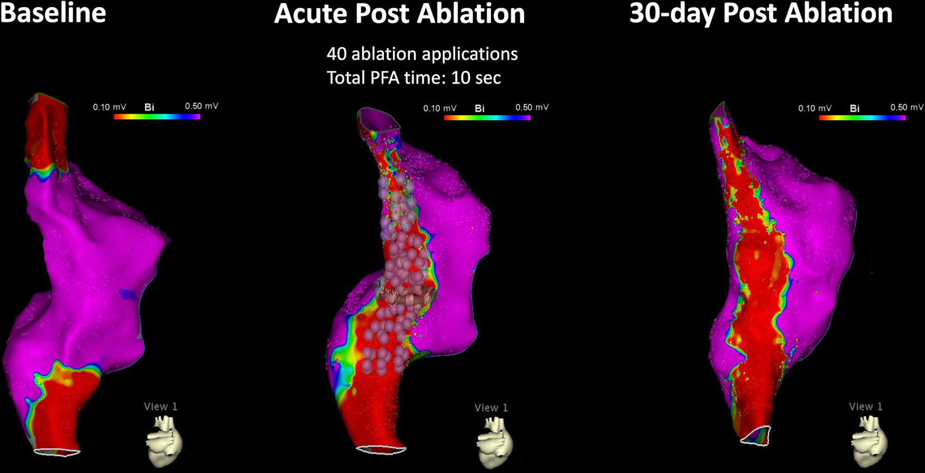
Methods: A 7.5F bidirectional circular catheter with 10 electrodes and variable expansion was designed for PFA (biphasic, 1800 Volts). This study included a total of 16 swine used to investigate the following 3 experimental aims: Aim 1 examined the feasibility to create a right atrial ablation line of block from the superior vena cava to the inferior vena cava. Aim 2 examined the effect of PFA on lesion maturation including durability after a 30-day survival period. Aim 3 examined the effect of high-intensity PFA (10 applications) on esophageal and phrenic nerve tissue in comparison to normal intensity radiofrequency ablation (1-2 applications). Histopathologic analysis of all cardiac, esophageal, and phrenic nerve tissue was performed.

Results: Acute line of block was achieved in 12/12 swine (100%) and required a total PFA time of 14 seconds (interquartile range [IQR], 9-24.5) per line. Ablation line durability after 28±3 days was maintained in 11/12 (91.7%) swine. PFA resulted in transmural lesions in 179/183 (97.8%) sections and a median lesion width of 14.2 mm. High-intensity PFA (9 [IQR, 8-14] application) had no effect on the esophagus while standard intensity radiofrequency ablation (1.5 [IQR, 1-2] applications) resulted in deep esophageal tissue injury involving the muscularis propria and adventitia layers. High-intensity PFA (16 [IQR, 10-28] applications) has no effect on phrenic nerve function and structure while standard dose radiofrequency ablation (1.5 [IQR, 1-2] applications) resulted in acute phrenic nerve paralysis.

Conclusions: In this preclinical model, a multielectrode circular catheter and multichannel generator produced durable atrial lesions with lower vulnerability to esophageal or phrenic nerve damage.

Keywords: adventitia; atrial fibrillation; catheter ablation; electrodes; paralysis.


Conflict of interest statement

Disclosures: Dr. Anter has received research grants and consultation fees from Biosense Webster, Inc. He also receives research grants and consultation fees from Affera Inc., Boston Scientific, Itamar Medical and Phillips Health. Drs, Brem, Zilberman, and Datta are employees at Biosense Webster. All other authors have no conflicts of interest relevant to this study.

Figures

Figure 1:
Figure 1:
Description of the Catheter. The investigational catheter is a circular multielectrode catheter with variable expansion ranging from 25 to 35mm. It includes 10 electrodes capable of pulsed field ablation in bipolar configuration. A magnetic sensor allows visualization by the electroanatomical mapping system.
Figure 2:
Figure 2:
Feasibility and Durability of a Right Atrial Ablation Line. The experimental model included a baseline activation map of the right atrium during pacing from the lateral tricuspid annulus (left panel). Pulsed field ablation was then performed along the posterior wall from the superior to the inferior vena cava. The presence or absence of activation line of block was examined by repeat mapping of the chamber during pacing from a similar location, immediately (middle panel) and 30-days after ablation (right panel). Note the presence of a durable block.
Figure 3:
Figure 3:
A Time-Dependent Effect of PFA on Voltage Amplitude. This figure shows the bipolar voltage map of the swine depicted in Figure 2. Note a continuous and durable line of voltage amplitude ≤0.1mV corresponding to the location of the ablation tags (each pulsed field ablation application is represented by 3 tags- indicating the participating electrodes).
Figure 4:
Figure 4:
Electrogram Attenuation with Pulsed Field Ablation. Pulsed field ablation was applied between electrodes 4–6 for a duration of ~250 microseconds. During the application period, electrograms (EGMs) are saturated. However, immediately afterward, significant EGM attenuation is observed.
Figure 5:
Figure 5:
Histopathological Examination of Pulsed Field Ablation in the Atrium. The left panel shows an example of a pathological specimen of a posterior ablation line demarcated by the dashed lines. The right panel shows histological slides corresponding to the indicated level. The upper panel shows a lower magnification slide in Mason’s Trichrome stain where a blue color indicates collagen. Note replacement of the posterior wall by collagen and fat. The lower panels show higher magnification in mason trichome (left) and Hematoxylins and Eosin (right). Note, the preserved nerve bundles within the ablation lesion. E=endocardium, N=nerve bundle, PW=posterior wall.
Figure 6:
Figure 6:
The Effect of Pulsed Field and Radiofrequency Ablation on the Esophagus. The close proximity between the right inferior pulmonary vein (RIPV) and the esophagus in swine served as the basis for comparing the effect of pulsed field ablation (PFA) and radiofrequency ablation (RFA) on the esophagus. The left atrium and RIPV are indicated by the green colored chamber, the esophagus by the brown colored tube, and the right atrium by the gray colored chamber. Insertion of the circular ablation catheter into the RIPV resulted in expansion of the RIPV and juxtaposition between the RIPV and the esophagus (median distance of 1.8mm). PFA had no effect on the esophagus following a 30-day survival period while RFA resulted in acute injury to the muscularis propria.
Figure 7:
Figure 7:
The Effect of Pulsed Field and Radiofrequency Ablation on the Phrenic Nerve. The vulnerability of the phrenic nerve to pulsed field ablation (PFA) and radiofrequency ablation (RFA) was examined by ablation in the lateral right atrium overlying the right phrenic nerve (marked by the yellow tags). Multiple PFA applications (purple tags) had no effect on phrenic nerve function or structure. In contrast, 2 RFA applications (red tags) resulted in acute phrenic nerve paralysis. The middle and right panels show the right phrenic nerve with localized swelling and whiting corresponding to sites of RFA.

References

    1. Chang DC, Reese TS. Changes in membrane structure induced by electroporation as revealed by rapid-freezing electron microscopy. Biophys J. 1990;58:1–12. - PMC - PubMed
    1. Neven K, van Driel V, van Wessel H, van Es R, du Pre B, Doevendans PA, Wittkampf F. Safety and feasibility of closed chest epicardial catheter ablation using electroporation. Circ Arrhythm Electrophysiol. 2014;7:913–9. - PubMed
    1. van Driel VJ, Neven K, van Wessel H, Vink A, Doevendans PA, Wittkampf FH. Low vulnerability of the right phrenic nerve to electroporation ablation. Heart Rhythm. 2015;12:1838–44. - PubMed
    1. Neven K, van Es R, van Driel V, van Wessel H, Fidder H, Vink A, Doevendans P, Wittkampf F. Acute and Long-Term Effects of Full-Power Electroporation Ablation Directly on the Porcine Esophagus. Circ Arrhythm Electrophysiol. 2017;10:e004672. - PubMed
    1. Bradley CJ, Haines DE. Pulsed field ablation for pulmonary vein isolation in the treatment of atrial fibrillation. J Cardiovasc Electrophysiol. 2020;31:2136–2147. - PubMed

Publication types