Accuracy of apparent diffusion coefficient in differentiation of glioblastoma from metastasis
- PMID: 33417503
- PMCID: PMC8165902
- DOI: 10.1177/1971400920983678
Accuracy of apparent diffusion coefficient in differentiation of glioblastoma from metastasis
Abstract
Background: Brain metastasis and glioblastoma multiforme are two of the most common malignant brain neoplasms. There are many difficulties in distinguishing these diseases from each other.
Purpose: The purpose of this study was to determine whether the mean apparent diffusion coefficient and absolute standard deviation derived from apparent diffusion coefficient measurements can be used to differentiate glioblastoma multiforme from brain metastasis based on cellularity levels.
Material and methods: Magnetic resonance images of 34 patients with histologically verified brain tumors were evaluated retrospectively. Apparent diffusion coefficient and standard deviation values were measured in the enhancing tumor, peritumoral region, and contralateral healthy white matter. Then, to determine whether there was a statistical difference between brain metastasis and glioblastoma multiforme, we analyzed different variables between the two groups.
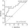
Results: Neither mean apparent diffusion coefficient values and ratios nor standard deviation values and ratios were significantly different between glioblastoma multiforme and brain metastasis. Receiver operating characteristic curve analysis of the logistic model with backward stepwise feature selection yielded an area under the curve of 0.77, a specificity of 84%, a sensitivity of 67%, a positive predictive value of 83.33%, and a negative predictive value of 78.26% for distinguishing between glioblastoma multiforme and brain metastasis. The absolute standard deviation and standard deviation ratios were significantly higher in the peritumoral edema compared to the tumor region in each case.
Conclusion: Apparent diffusion coefficient values and ratios, as well as standard deviation values and ratios in peritumoral edema, cannot be used to differentiate edema with infiltration of tumor cells from vasogenic edema. However, standard deviation values could successfully characterize areas of peritumoral edema from the tumoral region in each case.
Keywords: Apparent diffusion coefficient; brain metastasis; diffusion weighted imaging; glioblastoma multiforme.
Figures


Similar articles
-
Diagnostic value of peritumoral minimum apparent diffusion coefficient for differentiation of glioblastoma multiforme from solitary metastatic lesions.AJR Am J Roentgenol. 2011 Jan;196(1):71-6. doi: 10.2214/AJR.10.4752. AJR Am J Roentgenol. 2011. PMID: 21178049
-
Quantitative apparent diffusion coefficients in the characterization of brain tumors and associated peritumoral edema.Acta Radiol. 2009 Jul;50(6):682-9. doi: 10.1080/02841850902933123. Acta Radiol. 2009. PMID: 19449234
-
Nonenhancing peritumoral hyperintense lesion on diffusion-weighted imaging in glioblastoma: a novel diagnostic and specific prognostic indicator.J Neurosurg. 2018 Mar;128(3):667-678. doi: 10.3171/2016.10.JNS161694. Epub 2017 Mar 31. J Neurosurg. 2018. PMID: 28362236
-
Gradient of apparent diffusion coefficient values in peritumoral edema helps in differentiation of glioblastoma from solitary metastatic lesions.AJR Am J Roentgenol. 2014 Jul;203(1):163-9. doi: 10.2214/AJR.13.11186. AJR Am J Roentgenol. 2014. PMID: 24951211
-
Diffusion imaging of brain tumors.NMR Biomed. 2010 Aug;23(7):849-64. doi: 10.1002/nbm.1544. NMR Biomed. 2010. PMID: 20886568 Free PMC article. Review.
Cited by
-
Application of magnetic resonance imaging-related techniques in the diagnosis of sepsis-associated encephalopathy: present status and prospect.Front Neurosci. 2023 May 25;17:1152630. doi: 10.3389/fnins.2023.1152630. eCollection 2023. Front Neurosci. 2023. PMID: 37304016 Free PMC article. Review.
-
Advances in the application of neuroinflammatory molecular imaging in brain malignancies.Front Immunol. 2023 Jul 18;14:1211900. doi: 10.3389/fimmu.2023.1211900. eCollection 2023. Front Immunol. 2023. PMID: 37533851 Free PMC article. Review.
-
Neuroinflammation and immunoregulation in glioblastoma and brain metastases: Recent developments in imaging approaches.Clin Exp Immunol. 2021 Dec;206(3):314-324. doi: 10.1111/cei.13668. Epub 2021 Oct 8. Clin Exp Immunol. 2021. PMID: 34591980 Free PMC article. Review.
-
Differentiation of multiple brain metastases and glioblastoma with multiple foci using MRI criteria.BMC Med Imaging. 2024 Jan 2;24(1):3. doi: 10.1186/s12880-023-01183-3. BMC Med Imaging. 2024. PMID: 38166651 Free PMC article.
References
-
- Ostrom QT, Wright CH, Barnholtz-Sloan JS. Brain metastases: Epidemiology. In: Schiff D and Van den Bent MJ (eds.) Handbook of clinical neurology, Vol. 149. Elsevier, 2018, pp.27–42. Available at: https://www.sciencedirect.com/science/article/pii/B9780128111611000025 - PubMed
-
- Bucholtz JD. Central nervous system metastases. In: Ramakrishna R, Magge RS, Baaj AA and Knisely JPS (eds.) Cham: Springer International Publishing, 2020; pp.53–67.
-
- Lemercier P, Maya SP, Patrie JT, et al.. Gradient of apparent diffusion coefficient values in peritumoral edema helps in differentiation of glioblastoma from solitary metastatic lesions. Am J Roentgenol 2014; 203: 163–169. - PubMed
MeSH terms
LinkOut - more resources
Full Text Sources
Other Literature Sources
Medical

