. 2021 Jan 8;16(1):e0244648.
doi: 10.1371/journal.pone.0244648. eCollection 2021.

Transcriptomic analysis of the honey bee (Apis mellifera) queen spermathecae reveals genes that may be involved in sperm storage after mating

Affiliations

Transcriptomic analysis of the honey bee (Apis mellifera) queen spermathecae reveals genes that may be involved in sperm storage after mating

Juliana Rangel et al. PLoS One. .

Abstract

Honey bee (Apis mellifera) queens have a remarkable organ, the spermatheca, which successfully stores sperm for years after a virgin queen mates. This study uniquely characterized and quantified the transcriptomes of the spermathecae from mated and virgin honey bee queens via RNA sequencing to identify differences in mRNA levels based on a queen's mating status. The transcriptome of drone semen was analyzed for comparison. Samples from three individual bees were independently analyzed for mated queen spermathecae and virgin queen spermathecae, and three pools of semen from ten drones each were collected from three separate colonies. In total, the expression of 11,233 genes was identified in mated queen spermathecae, 10,521 in virgin queen spermathecae, and 10,407 in drone semen. Using a cutoff log2 fold-change value of 2.0, we identified 212 differentially expressed genes between mated and virgin spermathecal queen tissues: 129 (1.4% of total) were up-regulated and 83 (0.9% of total) were down-regulated in mated queen spermathecae. Three genes in mated queen spermathecae, three genes in virgin queen spermathecae and four genes in drone semen that were more highly expressed in those tissues from the RNA sequencing data were further validated by real time quantitative PCR. Among others, expression of Kielin/chordin-like and Trehalase mRNAs was highest in the spermathecae of mated queens compared to virgin queen spermathecae and drone semen. Expression of the mRNA encoding Alpha glucosidase 2 was higher in the spermathecae of virgin queens. Finally, expression of Facilitated trehalose transporter 1 mRNA was greatest in drone semen. This is the first characterization of gene expression in the spermathecae of honey bee queens revealing the alterations in mRNA levels within them after mating. Future studies will extend to other reproductive tissues with the purpose of relating levels of specific mRNAs to the functional competence of honey bee queens and the colonies they head.


Conflict of interest statement

The authors have declared that no competing interests exist.

Figures

Fig 1
Fig 1
Pictures depicting a) a honey bee queen; b) a virgin queen’s spermatheca with the tracheal net removed (left), a tracheal net (middle) and a mated queen’s spermatheca (diameter = ∼1 mm) filled with semen and the tracheal net removed (right) on a fingertip; c) an adult drone; and d) a fully everted endophallus with semen expressed at the end of the bulb (diameter of the semen droplet = ∼0.5 mm). Photos credits: a and c) Pixabay.com; b and d) Sue Cobey.
Fig 2
Fig 2
Volcano plots displaying differentially expressed genes (red-orange dots) between a) the spermatheca of mated and virgin honey bee queens, and b) the spermatheca of mated queens and drone semen. Each dot represents one gene. The black dots represent genes that were not differentially expressed (P < 0.01 and |log2 (Fold-change)| ≥ 2).
Fig 3
Fig 3. Heat map showing the relative expression levels of nine genes selected from RNA sequencing data for confirmation of differential expression by real time quantitative PCR (RT-qPCR).
The genes are identified by their BeeBase gene identifier. The tissues used (n = 3 biological replicates per tissue type) were spermathecae from mated queens (“Mated”), spermathecae from virgin queens (“Virgin”) and drone semen (“Drone”). The color index above indicates genes that were expressed at relatively low levels (blue) or at high levels (red) in each row. Heat maps were generated using Morpheus (https://software.broadinstitute.org/morpheus/).
Fig 4
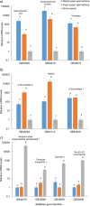
Fig 4. Real time quantitative PCR (RT-qPCR) confirmation revealed the relative expression levels of genes (identified by their BeeBase gene identifier) that were more highly expressed in mated queen spermatheca (blue bars), virgin queen spermatheca (orange bars), or drone semen (grey bars).
Of the selected genes, a) two of three genes were most highly expressed in the mated queen spermatheca; b) two of three genes were most highly expressed in the virgin queen spermatheca; and c) four genes were most highly expressed in drone semen. Different letters above each bar represent the tissue for which gene expression was significantly different from that in the other tissues (P < 0.05). Linear values are presented after normalizing to the EF1aF2 mRNA. Protein names are listed above the bars.

References

    1. Heifetz Y, Rivlin PK. Beyond the mouse model: Using Drosophila as a model for sperm interaction with the female reproductive tract. Theriogenology 2010;73:723–739. 10.1016/j.theriogenology.2009.11.001 - DOI - PubMed
    1. Gotoh A, Shigenobu S, Yamaguchi K, Kobayashi S, Ito F, Tsuji K. Transcriptome profiling of the spermatheca identifies genes potentially involved in the long-term sperm storage of ant queens. Sci Rep 2017;7:5972 10.1038/s41598-017-05818-8 - DOI - PMC - PubMed
    1. Orr TJ, Zuk M. Sperm storage. Curr Biol 2012;22:R8–R10. 10.1016/j.cub.2011.11.003 - DOI - PubMed
    1. Pascini TV, Ramalho-Ortigão M, Ribeiro JM, Jacobs-Lorena M, Ferreira Martins G. Transcriptional profiling and physiological roles of Aedes aegypti spermathecal-related genes. BMC Genomics 2020;21:143 10.1186/s12864-020-6543-y - DOI - PMC - PubMed
    1. Pitnick S, Markow T, Spicer GS. Evolution of multiple kinds of female sperm-storage organs in Drosophila. Evolution 1999;53:1804–1822. 10.1111/j.1558-5646.1999.tb04564.x - DOI - PubMed

Publication types

LinkOut - more resources