Review
. 2021 Jan 7;184(1):18-32.
doi: 10.1016/j.cell.2020.12.002.

Bioprinting for the Biologist

Affiliations
Review

Bioprinting for the Biologist

Andrew C Daly et al. Cell. .

Abstract

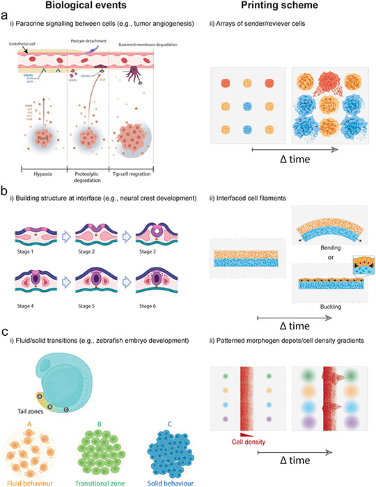
Building tissues from scratch to explore entirely new cell configurations could revolutionize fundamental understanding in biology. Bioprinting is an emerging technology to do this. Although typically applied to engineer tissues for therapeutic tissue repair or drug screening, there are many opportunities for bioprinting within biology, such as for exploring cellular crosstalk or cellular morphogenesis. The overall goals of this Primer are to provide an overview of bioprinting with the biologist in mind, outline the steps in extrusion bioprinting (the most widely used and accessible technology), and discuss alternative bioprinting technologies and future opportunities for bioprinting in biology.


Figures

Figure 1.
Figure 1.. Extrusion bioprinting process.
In extrusion bioprinting, a bioink (formulation of cells, often with a material) is deposited from a printer nozzle either onto a surface (top) or within a suspension bath (bottom) with a user-defined pattern. There are numerous commercially available bioprinters and biomaterials for use in bioinks that are making bioprinting accessible to many users.
Figure 2.
Figure 2.. The bioprinting experimental workflow.
This consists of three general parts: (A) planning (e.g., creating a print design and defining the bioink and biomaterial ink to be used), (B) printing the construct, and (C) processing the printed construct over time with cell culture and identifying appropriate analytical outcomes.
Figure 3.
Figure 3.. Bioprinted models of tissue development and disease.
(a) i) Angiogenic sprouting - Schematic of 3D printed microchannels within cell degradable hydrogels where the left channel is seeded with endothelial cells and the right channel is perfused with angiogenic factors, and ii) endothelial cell sprouting from the vascular channel towards the VEGF gradient over 3 days of culture. (Song et al., 2018) iii) Tissue buckling - Schematic demonstrating embedded 3D printing of collagen filaments containing fibroblasts into a granular yield stress media (i.e., suspension bath), and fibroblast contraction of the collagen matrix inside the printed filament, and iv) contraction and buckling of the collagen filament over 24 hours of culture as a function of the filament aspect ratio (length/diameter). (Morley et al., 2019) (b) i) Glioblastoma model – Schematic and image of extrusion bioprinting of a mini-brain model containing compartmentalized regions of glioblastoma cells and macrophages to study the role of macrophages in glioblastoma progression. (Heinrich et al., 2019) ii) Renal proximal tubule model – Schematic demonstrating 3D printing of a sacrificial pluronic ink to generate convoluted perfusable channels inside a gelatin/fibrin matrix, and iv) seeding of the microchannels with proximal tubule epithelial cells (PTECS) and glomerular microvascular endothelial cells (GMECs) to generate parallel vascular and renal epithelial channels to study solute renal reabsorption. (Lin et al., 2019)
Figure 4.
Figure 4.. Advanced bioprinting technologies.
(a) i) DLP lithography bioprinting process where light is spatially projected onto a cell laden bioresin using a digital micromirror device to create a liver lobule construct (green regions contain iPSC-hepatocytes, and red regions contain endothelial cells & adipose-derived stem cells. Scalebars 500μm. (Ma et al., 2016) ii) DLP bioprinting of an alveolar lung model containing a central mechanically ventilated air sac surrounded by vascular channels perfused with red blood cells, and demonstration of gaseous exchange through measurement of reoxygenation of the red blood cell population (green line) following oxygenation of the air sac (blue line). Scalebars 1mm. (Grigoryan et al., 2019) iii) Experimental setup for volumetric bioprinting including laser input followed by DLP projection modulation of light onto a rotating platform containing the bioresin. Image of bioprinted human ear model created using a cell laden GelMA bioresin, total printing time 22.7 seconds. Scalebar 2mm. (Bernal et al., 2019; Loterie et al., 2020) (b) i) Kenzan bioprinting method where cell spheroids are aspirated and then skewered onto metal needles for fusion into 3D constructs. (Moldovan et al., 2016) ii) Aspiration-assisted bioprinting of spheroids (labelled red and green) onto fibrin hydrogels at different separation distances to study paracrine signaling and angiogenic sprouting. Scalebar 400μm. (Ayan et al., 2020)
Figure 5.
Figure 5.. Bioprinting approaches to biological questions in tissue development and homeostasis.
(a) To study interactions between endothelial and tumor cells in a highly controlled manner, spatial combinatorial patterning of engineered “sender” and “receiver” cell arrays could test hypotheses around diffusible biochemical signaling and their influence on cell phenotype and function (e.g., protein/gene expression, migration, proliferation). This could be achieved by patterning cell depots of distinct compositions at prescribed spacing within ECM hydrogels and then monitoring cell behaviour during culture. (b) To study biophysical morphogenesis in neural crest development strains at bioprinted tissue interfaces could be generated through internally generated cell-cell or cell-ECM forces to create dynamic changes in tissue shape. Bioprinting could be used to interface two filaments where differences in cell behaviour (e.g., contractility, proliferation) within filaments drive bending or buckling behaviours. (c) Gradients in growth factor concentrations could stimulate formation of fluid-like and solid-like domains to guide dynamic remodeling of bioprinted tissues, and to study microenvironmental conditions that drive tissue fluidity (i.e., interplay between cell-cell interactions, cell-ECM interactions, and morphogen presentation), such as during head-to-tail elongation in the zebrafish embryo. Models could be produced by patterning morphogen depots adjacent to filaments containing cell density gradients, allowing combinatorial screening of cell migration behaviour across conditions.

References

    1. Ahlfeld T, Doberenz F, Kilian D, Vater C, Korn P, Lauer G, Lode A, and Gelinsky M (2018). Bioprinting of mineralized constructs utilizing multichannel plotting of a self-setting calcium phosphate cement and a cell-laden bioink. Biofabrication 10, 045002. - PubMed
    1. Ahlfeld T, Guduric V, Duin S, Akkineni AR, Schütz K, Kilian D, Emmermacher J, Cubo-Mateo N, Dani S, Witzleben M.v., et al. (2020). Methylcellulose – a versatile printing material that enables biofabrication of tissue equivalents with high shape fidelity. Biomaterials science 8, 2102–2110. - PubMed
    1. Aisenbrey EA, and Murphy WL (2020). Synthetic alternatives to Matrigel. Nature Reviews Materials 5, 539–551. - PMC - PubMed
    1. Andrew DJ, and Ewald AJ (2010). Morphogenesis of epithelial tubes: Insights into tube formation, elongation, and elaboration. Dev Biol 341, 34–55. - PMC - PubMed
    1. Aw WY, Heck BW, Joyce B, and Devenport D (2016). Transient Tissue-Scale Deformation Coordinates Alignment of Planar Cell Polarity Junctions in the Mammalian Skin. Curr Biol 26, 2090–2100. - PMC - PubMed

Publication types