Review
. 2021 Mar 10:331:103-120.
doi: 10.1016/j.jconrel.2020.12.057. Epub 2021 Jan 6.

Breast tumor-on-chip models: From disease modeling to personalized drug screening

Affiliations
Review

Breast tumor-on-chip models: From disease modeling to personalized drug screening

Bano Subia et al. J Control Release. .

Abstract

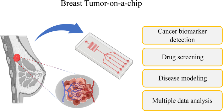
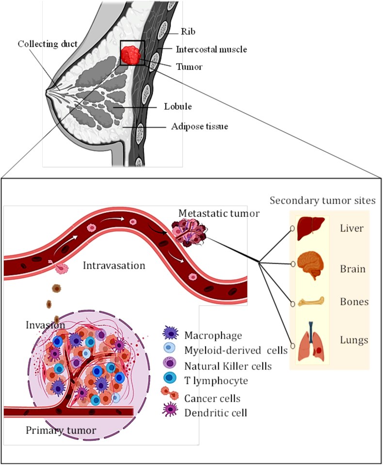
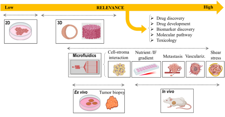
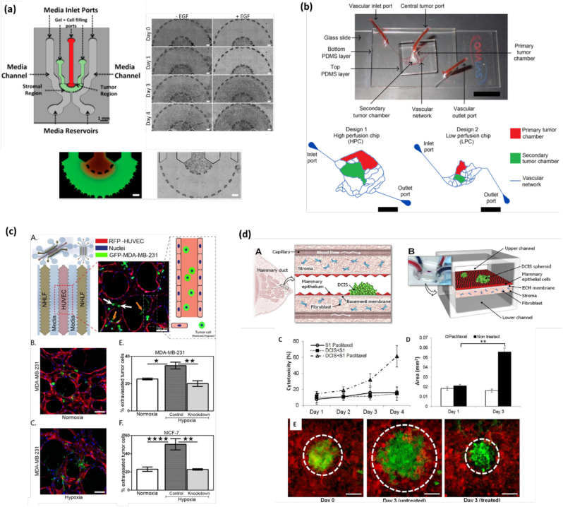
Breast cancer is one of the leading causes of mortality worldwide being the most common cancer among women. Despite the significant progress obtained during the past years in the understanding of breast cancer pathophysiology, women continue to die from it. Novel tools and technologies are needed to develop better diagnostic and therapeutic approaches, and to better understand the molecular and cellular players involved in the progression of this disease. Typical methods employed by the pharmaceutical industry and laboratories to investigate breast cancer etiology and evaluate the efficiency of new therapeutic compounds are still based on traditional tissue culture flasks and animal models, which have certain limitations. Recently, tumor-on-chip technology emerged as a new generation of in vitro disease model to investigate the physiopathology of tumors and predict the efficiency of drugs in a native-like microenvironment. These microfluidic systems reproduce the functional units and composition of human organs and tissues, and importantly, the rheological properties of the native scenario, enabling precise control over fluid flow or local gradients. Herein, we review the most recent works related to breast tumor-on-chip for disease modeling and drug screening applications. Finally, we critically discuss the future applications of this emerging technology in breast cancer therapeutics and drug development.

Keywords: Breast cancer; Drug screening; Industrial applications; Microfluidics; Tumor-on-chip.


Conflict of interest statement

SB, JA and GVC are employers of Elvesys. Elveflow is an Elvesys brand. Authors declare no other conflicts of interest.

Figures

Unlabelled Image
Graphical abstract
Fig. 1
Fig. 1
The tumor microenvironment (TME) and the cascade of breast cancer metastasis. Tumor dissemination is initiated by the uncontrolled growth of the tumor and the formation of angiogenesis, a process where new blood vessels are formed from the preexisting ones. These vessels are employed to provide nutrients and oxygen to the tumor. Next, metastatic cancer cells invade the surrounding TME and migrate directionally towards the microvasculature to invade it in a process known as intravasation. Then, these tumor cells travel through the blood vessels as circulating tumor cells (CTCs) to invade distant organs. Many of these CTCs are destroyed or damaged during the circulation due to their inability to transit through the capillaries. A few undamaged cells may extravasate and invade the parenchyma of a foreign tissue (e.g., liver, brain, bone, or lung). At the invading stage, cancer cells start proliferating forming a secondary tumor site. Therein, multiple immune cells, such as macrophages, natural killer cells, T lymphocytes and dendritic cells, reside in the tumor niche (Created using Biorender.com).
Fig. 2
Fig. 2
Overview of in vitro models for studying breast cancer physiopathology and for drug screening applications. The 2D tumor model is typically represented by a monolayer culture of cells; 3D tumor models (e.g., spheroids, cancer cells encapsulated within scaffolds/hydrogels, microcarriers, and others) can reproduce native cell-cell communication and cell-ECM interactions. Ex vivo (tumor biopsy) and in vivo models can be used for drug screening, drug discovery and development, biomarkers detection and to indentify molecular pathways involved in breast tumor. Microfluidic chip models can mimic the in vivo physiopathology of breast cancer, such as vasculature growth, gradient generation, interstitial flow, or shear stress. In addition, important events of the metastatic cascade can be easily reproduced and studied, such as tumor growth, invasion, intravasation, vasculature CTC transit, extravasation, or organ specificity. (Created using Biorender.com).
Fig. 3
Fig. 3
Human breast tumor-on-a-chip models. (a) Mimicking gradient generation on-chip. (Left) Microfluidic device for the study of breast cancer cell invasion into the 3D stroma. Bottom images show the spatial organization of cells encapsulated within a 3D matrix. (Right) Time-sequence for 4 days showing the invasion of EFG+ and EGF- SUM-159 breast cancer cells into the neighboring stroma. Reproduced with permission from [5] (Creative Commons Attribution 4.0 International License). (b) Mimicking fluid dynamics on chip. (Top) Tumor-mimetic microfluidic chip containing a realistic vascular network. (Bottom) Schematic representation of the vascular network, primary and secondary tumor chambers. Reproduced with permission from [105] (Creative Commons Attribution 4.0 International License). (c) Mimicking hypoxia effect on-chip. (A) Microfluidic chip showing the distribution of NHLF, HUVEC, and invasive GFP-MDA-MB-231 breast cancer cells. (B—C) Immunofluorescence image under normoxia (B) and hypoxia (C) conditions. (E-F) Quantification of the % of extravasated tumor MDA-MB-231 (E) and MCF7 (F) cells for all conditions. Reproduced with permission from [107] (Creative Commons Attribution 4.0 International License). (d) Mimicking tumor-stroma interactions on-chip. (A) The Ductal caricinoma in situ (DCIS) is embedded in a mammary duct consisting of the mammary epithelium and a basement membrane surrounded by stromal tissue (fibroblasts). (B) The microarchitecture of the DCIS and the surrounding tissue layers is reproduced in the breast cancer-on-a-chip microdevice comprised of the upper and lower cell culture chambers separated by an ECM-derived porous membrane. (C) cells are treated with paclitaxel from the basal side to simulate intravenous administration. (D) Paclitaxel treatment prevents growth of DCIS spheroids (white). (E) Fluorescence micrographs of DCIS spheroids at day 0 (left), day 3 without paclitaxel (middle), and 3 days with paclitaxel treatment (right). Reproduced with permission from [16] (Creative Commons Attribution 4.0 International License; CC BY 4.0).
Fig. 4
Fig. 4
Overview of tumor therapy approach through microfluidic technology for drug screening and breast tumor marker detection and quantification. (a) Schematic representation of microfluidic setup. used for the sorting of circulatory tumor cells and breast tumor specific biomarkers from solid tumor and patient-derived samples . A A pressure controller and a flow sensor (MFS) are used to create a precise interstitial fluidic pressure and flow speed, similar to the perfused native cancerous tissue. It can be used for biomarker detection and drug treatment. (b) Schematic representation and image of the microfluidic chip for exosomes capture and detection (A and B). (c) (A) Quantification of EpCAM-positive exosomes from breast cancer cell lines from control, normal fibroblast, MCF7 and MDA-MB-231 culture medium. (B) Quantification of captured EpCAM-positive exosomes from plasma samples of breast cancer patients and healthy controls, Reproduced with permission from [146] (Creative Commons Attribution 4.0 International License). (d) Screening studies of breast cancer cell lines in 2D and 3D microfluidic culture (bottom left). a, HCC-1937 (TNBCs) were seeded in 3D OrganoPlate® and 2D tissue culture plates. Paclitaxel and olaparib drugs were added after 72 h. b, Similarly, MDA-MB-231 and MDA-MB-453 cells were seeded and exposed to cisplatin at various concentrations and cellular viability was quantified. Reproduced with permission from [21] (Creative Commons Attribution 4.0 International License).
Fig. 5
Fig. 5
Workflow for a drug screening setup with breast tumor biopsy sample. Blood samples are transferred to the microfluidic chip and incubated with chemical compound libraries. The most efficient drug combination should be determined based on different breast cancer biomarker and phenotypic data. The effect of compounds on blood samples is measured using a variety of cell-based readout and pharmacokinetics/pharmacodynamics (PK/PD) assays. These detection assays can be used for monitoring the cell viability/toxicity and cellular function by the measurement of homogenous changes in absorbance, fluorescence- or luminescence-based gene reporter assays. PK/PD can provide information to simulate drug responses in the human body.
Fig. 6
Fig. 6
Overview of the functional and genetic cancer patient stratification and disease diagnosis approach using microfluidics. Tumor biopsies can be used to determine first line of treatment strategy based on their genetic and phenotypic data. The droplet-based microfluidic technology can be used to isolate CTCs from patients to monitor the disease state. The mass accumulation rate can be monitored through a series of suspended microchannel resonators to allow drug susceptibilities of patient derived tumor cells or CTCs. In combination, genotypic testing provides the information to predict the effect of potent drugs from patients' genotypes. These data can be used by clinicians for the selection of drug for precise treatment therapy. Reproduced with permission from ref. [151] (Creative Commons Attribution 4.0 International License (CC BY 4.0)).

Similar articles

Cited by

References

    1. Wang X., Wang N., Zhong L., Wang S., Zheng Y., Yang B., Zhang J., Lin Y., Wang Z. Mol. Psychiatry. 2020;25:3186–3197. doi: 10.1038/s41380-020-00865-6. - DOI - PMC - PubMed
    1. Aleskandarany M.A., Vandenberghe M.E., Marchiò C., Ellis I.O., Sapino A., Rakha E.A. Pathobiology. 2018;85:23–34. doi: 10.1159/000477851. - DOI - PubMed
    1. Feng Y., Spezia M., Huang S., Yuan C., Zeng Z., Zhang L., Ji X. Genes Dis. 2018;5(2):77–106. doi: 10.1016/j.gendis.2018.05.001. - DOI - PMC - PubMed
    1. Brancato V., Gioiella F., Imparato G., Guarnieri D., Urciuolo F., Netti P.A. Acta Biomater. 2018 doi: 10.1016/j.actbio.2018.05.055. - DOI - PubMed
    1. Truong D., Puleo J., Llave A., Mouneimne G., Kamm R.D., Nikkhah M. Sci. Rep. 2016 doi: 10.1038/srep34094. - DOI - PMC - PubMed

Publication types