. 2021 Jan 6;22(2):477.
doi: 10.3390/ijms22020477.

Generation of a Novel Mesothelin-Targeted Oncolytic Herpes Virus and Implemented Strategies for Manufacturing

Affiliations

Generation of a Novel Mesothelin-Targeted Oncolytic Herpes Virus and Implemented Strategies for Manufacturing

Guendalina Froechlich et al. Int J Mol Sci. .

Abstract

Background: HER2-based retargeted viruses are in advanced phases of preclinical development of breast cancer models. Mesothelin (MSLN) is a cell-surface tumor antigen expressed in different subtypes of breast and non-breast cancer. Its recent identification as a marker of some triple-negative breast tumors renders it an attractive target, presently investigated in clinical trials employing antibody drug conjugates and CAR-T cells. The availability of MSLN-retargeted oncolytic viruses may complement the current immunotherapeutic panel of biological drugs against HER2-negative breast and non-breast tumors.

Methods: A fully virulent, tumor-targeted oncolytic Herpes simplex virus-1 (MSLN-THV) with a selectivity for mesothelin-expressing cancer cells was generated. Recombineering technology was used to replace an essential moiety of the viral glycoprotein D with antibody fragments derived from clinically validated MSLN monoclonal antibodies, and to allow IL12 cargo expression in infected cells. Panels of breast and female reproductive system cell lines were used to verify the oncolytic potential of the viral constructs. A platform for production of the retargeted viruses was developed in HEK 293 cells, providing stable expression of a suitable chimeric receptor.

Results: We demonstrated the selectivity of viral infection and cytotoxicity by MSLN-retargeted viruses in a panel of mesothelin-positive cancer cells, originating from breast and female reproductive system tumors. We also developed a second-generation oncolytic MSLN-THV, encoding IL12, to enhance the immunotherapeutic potential of the viral backbone. A non-tumor cell line expressing a chimeric MSLN/Nectin-1 receptor, de-sensitized from antiviral responses by genetic inactivation of the Stimulator of Interferon Genes (STING)-dependent pathway was engineered, to optimize viral yields.

Conclusions: Our proof-of-concept study proposes MSLN-retargeted herpesviruses as potential cancer immunotherapeutics for assessments in preclinical models of MSLN-positive tumors, complementing the available panel of oncolytic viruses to HER2-negative breast tumors.

Keywords: MSLN; malignant mesothelioma; oncolytic virus; targeted therapy; triple negative breast cancer.


Conflict of interest statement

The authors Alfredo Nicosia and Elisa Scarselli are founders and shareholders of Nouscom S.R.L. Emanuele Sasso, Gabriella Cotugno, Anna Morena D’Alise, Armin Lahm are employees of Nouscom S.R.L. All the other authors declare no competing interests.

Figures
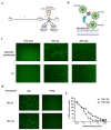
Figure 1
Figure 1
Generation of Herpes simplex type 1 (HSV-1)-based oncolytic viruses retargeted to Mesothelin (MSLN). (a) Three targeted Herpes viruses (THVs) were generated by substitution of the essential moiety of viral glycoprotein D (aa 6 to 38) with three different antibody fragments recognizing MSLN: SD1 (orange), SS (light blue), and its affinity matured form SS1 (green) (b) targeting two different domains of MSLN. (c) THV-SS, THV-SS1, and THV-SD1 Bacterial Artificial Chromosome (BAC) DNAs were transfected in SKOV3 cells to produce infectious viral particles (top panels); THV-SD1 failed to be recovered in first passage of infection (P1) (bottom panel). (d) Viral infection and spread were analyzed in presence of the monoclonal antibody Amatuximab (α-MSLN) used at saturating concentration (150 μg) to compete with THV-SS and THV-SS1 viruses. (e) The Amatuximab antibody was used at increasing concentrations and the number of eGFP-positive (infected) cells was determined. The statistical significance was calculated by Student’s t-test and resulted p < 0.05 in the range of Amatuximab concentration between 0.1 and 50 μg/mL.
Figure 2
Figure 2
Soluble MSLN does not interfere with virus spread and mIL12 production. (a) Secreted soluble MSLN in supernatant of SKOV3 cell line was quantified by ELISA assay up to 120 h post seed. Dashed lines define the range of soluble mesothelin (SMRP) concentration in tumor patients. The statistical significance was calculated by Student’s t-test and resulted p < 0.05 * comparing 24 and 48 h, p < 0.005 ** comparing 48 and 72 h/72 and 96 h/96 and 120 h. (b) The same conditioned media described in panel A were used to infect fresh SKOV3 cells with THV_SS (light blue) and THV_SS1 (green) resulting in limited alteration of viral entry at the highest SMRP concentrations. The statistical significance was calculated by Student’s t-test and resulted not significant (NS) comparing THV_SS and THV_SS1 at each time point and p < 0.05 * comparing both THV_SS and THV_SS1 at 0 and 60 ng/mL. (c) Schematic cartoon of second generation THV_SS1 encoding mIL-12 (THV_SS1-IL12); the mIL-12 expression cassette was inserted into the intergenic region US1/US2. (d) Production of mIL-12 cytokine quantified by ELISA assay from 48 to 96 h.
Figure 3
Figure 3
Cytopathic effect of THV_SS1 is tightly dependent on MSLN positive cells. (a) HEK293 cells (MSLN-) were infected from wild-type virus R-LM55, but not from THV_SS1 virus, confirming the stringency of the retargeting. (b) HEK293 cells were stably transduced with mesothelin protein and were efficiently infected (fluorescent positive cells) at different MOIs (MOI from 0.001 to 1) by THV_SS1, up to 120 h post-infection. Bright-field images show the cytotoxic effect of THV_SS1 virus (round detached cells). (c) Mesothelin retargeting does not impact on viral replication in proficient cells, as THV_SS1 and parental R-LM55 viruses replicate equally well in HEK293-MSLN cell line. (d) The cytotoxic effect of R-LM55 and THV_SS1 in HEK293 and HEK293-MSLN was assessed by Alamar Blue. After 6 days from infection, R-LM55 reached 70% of cytopathic effect in both HEK293 cells (+/−MSLN), while THV_SS1 reached 70% of cytopathic infect only in HEK293-MSLN and maintains 100% of live cells in wild-type HEK293 cells.
Figure 3
Figure 3
Cytopathic effect of THV_SS1 is tightly dependent on MSLN positive cells. (a) HEK293 cells (MSLN-) were infected from wild-type virus R-LM55, but not from THV_SS1 virus, confirming the stringency of the retargeting. (b) HEK293 cells were stably transduced with mesothelin protein and were efficiently infected (fluorescent positive cells) at different MOIs (MOI from 0.001 to 1) by THV_SS1, up to 120 h post-infection. Bright-field images show the cytotoxic effect of THV_SS1 virus (round detached cells). (c) Mesothelin retargeting does not impact on viral replication in proficient cells, as THV_SS1 and parental R-LM55 viruses replicate equally well in HEK293-MSLN cell line. (d) The cytotoxic effect of R-LM55 and THV_SS1 in HEK293 and HEK293-MSLN was assessed by Alamar Blue. After 6 days from infection, R-LM55 reached 70% of cytopathic effect in both HEK293 cells (+/−MSLN), while THV_SS1 reached 70% of cytopathic infect only in HEK293-MSLN and maintains 100% of live cells in wild-type HEK293 cells.
Figure 4
Figure 4
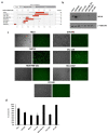
THV_SS1 infects breast and female reproductive system tumor cells in a MLSN-dependent manner. (a) Mesothelin expression assessed by GENEVESTIGATOR software by interrogating RNAseq repository of breast (CAL-120, HCC-1937, BT-549, MDA-MB-231) and female reproductive system (OVCAR3, HeLa, SKOV3) tumor cell lines. (b) MSLN expression in selected cell lines was evaluated by Western blot and gamma-tubulin was used as standard. (c) Infection and (d) cytotoxicity of THV_SS1 in tumor cell lines were assessed respectively by fluorescence/bright-field microscopy and trypan blue positive cells.
Figure 5
Figure 5
Generation of improved oncolytic virus producing cell line. (a) STING gene was knocked out in HEK293 cells as assessed by Western blot analysis; gamma-tubulin was used as standard. PCR screening confirmed the absence of eGFP and Cas9 residues in genomic DNA; Cas9/eGFP-encoding vector was used as positive control (C+) and genomic DNA from parental HEK293 cell was used as negative control (C−). (b) R-LM55 viral spread enhancement was demonstrated in HEK293 STING KO cell line compared to HEK293 WT cell line. (c,d) Enhancement viral yield was demonstrated on HEK293 STING KO vs. parental infected with R-LM55 and T-VEC viruses. The statistical significance was calculated by Student’s t-test comparing HEK293 STING KO and HEK 293 WT and resulted p < 0.005 ** and p < 0.05 *, respectively, for R-LM55 and T-VEC productivity.
Figure 6
Figure 6
Generation of oncolytic virus HEK293 STING KO producing cell line stably expressing an engineered non-secretable MSLN. (a) In the upper part is shown the chimeric MSLN-Nectin construct that consists of: Ig-κ signal peptide (white box), HA tag plus GA-based linker (blue box), the N-terminus 66 amino acid fragment of MSLN (E296-Q362) (red box), GSGA-based linker (green box), the C-terminus of Nectin-1 including the transmembrane and intracellular tail (M143-V517) (light-blue boxes). In the bottom part is shown a cartoon model that depicts the interaction of THV_SS1 with GPI-MSLN, SMRP, TM-MSLN proteins. Created by Biorender. (b) Western blot screening for HA tag to find TM-MSLN protein positive clones; clone H was chosen, and gamma-tubulin was used as standard. (c) HEK293_SKO_TM-MSLN cell line were efficiently infected by THV_SS1 at 0.005 MOI. (d) The secreted MSLN in SKOV3 and HEK293_SKO_TM-MSLN was quantified by ELISA assay. (e) The productivity of THV_SS1 was evaluated in SKOV3 at 0.01 MOI and in HEK293_SKO_TM-MSLN at different MOI (from 0.001 to 0.5). The statistical significance was calculated by Student’s t-test and resulted as not significant, comparing the different MOI in HEK293_SKO_TM-MSLN cells (0.5, 0.1, 0.05, 0.01, 0.005, 0.001 MOI). The difference between SKOV3 and HEK293_SKO_TM-MSLN resulted as significant (p < 0.05 *).

Similar articles

Cited by

References

    1. Sasso E., D’Alise A.M. New viral vector for infectious diseases and cancer. Semin. Immunol. 2020;50:101430. doi: 10.1016/j.smim.2020.101430. - DOI - PubMed
    1. Kaufman H.L., Kohlhapp F.J. Oncolytic viruses: A new class of immunotherapy drugs. Nat. Rev. Drug Discov. 2015;14:642–662. doi: 10.1038/nrd4663. Erratum in: Nat. Rev. Drug Discov.2016, 15, 660. - DOI - PMC - PubMed
    1. Bommareddy P.K., Shettigar M. Integrating oncolytic viruses in combination cancer immunotherapy. Nat. Rev. Immunol. 2018;18:498–513. doi: 10.1038/s41577-018-0014-6. Erratum in: Nat. Rev. Immunol.2018. - DOI - PubMed
    1. Sivanandam V., LaRocca C.J. Oncolytic Viruses and Immune Checkpoint Inhibition: The Best of Both Worlds. Mol. Ther. Oncolytics. 2019;13:93–106. doi: 10.1016/j.omto.2019.04.003. - DOI - PMC - PubMed
    1. Twumasi-Boateng K., Pettigrew J.L. Oncolytic viruses as engineering platforms for combination immunotherapy. Nat. Rev. Cancer. 2018;18:419–432. doi: 10.1038/s41568-018-0009-4. Erratum in: Nat. Rev. Cancer2018. - DOI - PubMed

LinkOut - more resources