Review
. 2021 Jan 6;13(2):170.
doi: 10.3390/cancers13020170.

The Role of Cellular Prion Protein in Promoting Stemness and Differentiation in Cancer

Affiliations
Review

The Role of Cellular Prion Protein in Promoting Stemness and Differentiation in Cancer

Larisa Ryskalin et al. Cancers (Basel). .

Abstract

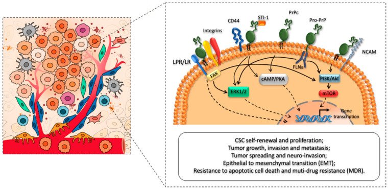
Cellular prion protein (PrPC) is seminal to modulate a variety of baseline cell functions to grant homeostasis. The classic role of such a protein was defined as a chaperone-like molecule being able to rescue cell survival. Nonetheless, PrPC also represents the precursor of the deleterious misfolded variant known as scrapie prion protein (PrPSc). This variant is detrimental in a variety of prion disorders. This multi-faceted role of PrP is greatly increased by recent findings showing how PrPC in its folded conformation may foster tumor progression by acting at multiple levels. The present review focuses on such a cancer-promoting effect. The manuscript analyzes recent findings on the occurrence of PrPC in various cancers and discusses the multiple effects, which sustain cancer progression. Within this frame, the effects of PrPC on stemness and differentiation are discussed. A special emphasis is provided on the spreading of PrPC and the epigenetic effects, which are induced in neighboring cells to activate cancer-related genes. These detrimental effects are further discussed in relation to the aberrancy of its physiological and beneficial role on cell homeostasis. A specific paragraph is dedicated to the role of PrPC beyond its effects in the biology of cancer to represent a potential biomarker in the follow up of patients following surgical resection.

Keywords: brain tumors; cancer stem cells; cellular prion protein; differentiation; peripheral tumors; self-renewal; tumorigenesis.


Conflict of interest statement

The authors declare no conflict of interest.

Figures

Figure 1
Figure 1
Cellular prion protein (PrPC) as a main regulator of CSCs phenotype, biology, and functioning. The left panel of the cartoon depicts the cross-talk between cancer stem cells (CSCs) and major cellular components (i.e., endothelial cells, fibroblasts, pericytes, macrophages, and T cells) of the tumor micro-environment (TME). The right panel schematizes the role of PrPC in promoting CSC self-renewal, proliferation, and migration. In addition, PrPC interacts with several receptors and cell surface proteins to modulate CSCs’ tumor-initiating and metastatic capacities, while promoting increased therapeutic resistance. In this way, it modulates tumor growth, survival, infiltration, and multi-drug resistance.
Figure 2
Figure 2
PrPC expression in human pancreatic tissues. (A) The figure reports representative pictures of PrPC immunohistochemistry of pancreatic ductal adenocarcinoma (PDAC) tissue (left panel) compared with control tissue (right panel). Immunoperoxidase shows PrPC-specific labelling in ductal cells (arrows) from a human tumor sample (left panel). Remarkably, the increase in PrPc expression occurs along with a marked loss of cellular architecture within pancreatic tumoral tissue, which features enlarged and irregularly shaped ducts within abundant extracellular matrix. In contrast, normal human pancreas (right panel) possesses a well-preserved architecture of both acinar cells and ductal system, along with a weak PrPC-staining in the ductal epithelial cells (original magnification 10×). (B) The figure reports a representative immunoblot for PrPC and the house keeping protein β-actin in control and PDAC tissues. (C) The figure reports representative western blotting comparing scrapie prion protein (PrPSc) and PrPC (with or without proteinase K) in control and PDAC tissues as measured in Table 1. Images were obtained by an author of the manuscript (M.A. Giambelluca). Original western blot images (Figures S1 and S2) and densitometry readings (Tables S1 and S2) were provided in Supplemental Materials.

References

    1. Prusiner S.B. Prions. Proc. Natl. Acad. Sci. USA. 1998;95:13363–13383. doi: 10.1073/pnas.95.23.13363. - DOI - PMC - PubMed
    1. Fornai F., Ferrucci M., Gesi M., Di Poggio A.B., Giorgi F.S., Biagioni F., Paparelli A. A hypothesis on prion disorders: Are infectious, inherited, and sporadic causes so distinct? Brain Res. Bull. 2006;69:95–100. doi: 10.1016/j.brainresbull.2005.12.005. - DOI - PubMed
    1. Prusiner S.B. Scrapie prions. Annu. Rev. Microbiol. 1989;43:345–374. doi: 10.1146/annurev.mi.43.100189.002021. - DOI - PubMed
    1. Prusiner S.B., Scott M.R., Dearmond S.J., Cohen F.E. Prion Protein Biology Review. Cell. 1998;93:337–348. doi: 10.1016/S0092-8674(00)81163-0. - DOI - PubMed
    1. Prusiner S.B. Molecular biology of prion diseases. Science. 1991;252:1515–1522. doi: 10.1126/science.1675487. - DOI - PubMed

LinkOut - more resources