Review
. 2021 Jan 6;10(1):61.
doi: 10.3390/antiox10010061.

Inflaming the Brain with Iron

Affiliations
Review

Inflaming the Brain with Iron

Pamela J Urrutia et al. Antioxidants (Basel). .

Abstract

Iron accumulation and neuroinflammation are pathological conditions found in several neurodegenerative diseases, including Alzheimer's disease (AD) and Parkinson's disease (PD). Iron and inflammation are intertwined in a bidirectional relationship, where iron modifies the inflammatory phenotype of microglia and infiltrating macrophages, and in turn, these cells secrete diffusible mediators that reshape neuronal iron homeostasis and regulate iron entry into the brain. Secreted inflammatory mediators include cytokines and reactive oxygen/nitrogen species (ROS/RNS), notably hepcidin and nitric oxide (·NO). Hepcidin is a small cationic peptide with a central role in regulating systemic iron homeostasis. Also present in the cerebrospinal fluid (CSF), hepcidin can reduce iron export from neurons and decreases iron entry through the blood-brain barrier (BBB) by binding to the iron exporter ferroportin 1 (Fpn1). Likewise, ·NO selectively converts cytosolic aconitase (c-aconitase) into the iron regulatory protein 1 (IRP1), which regulates cellular iron homeostasis through its binding to iron response elements (IRE) located in the mRNAs of iron-related proteins. Nitric oxide-activated IRP1 can impair cellular iron homeostasis during neuroinflammation, triggering iron accumulation, especially in the mitochondria, leading to neuronal death. In this review, we will summarize findings that connect neuroinflammation and iron accumulation, which support their causal association in the neurodegenerative processes observed in AD and PD.

Keywords: Alzheimer’s disease; Parkinson’s disease; hepcidin; iron; iron regulatory protein 1; neuroinflammation; nitric oxide; oxidative stress.


Conflict of interest statement

The authors declare no conflict of interest.

Figures

Figure 1
Figure 1
Regulatory mechanisms and functions of iron regulatory protein 1 (IRP1). The bifunctional protein c-aconitase/IRP1 is regulated by several mechanisms. C-aconitase is converted to IRP1 through two main processes: (i) Impaired Fe-S cluster biosynthesis, as a result of decreased mitochondrial iron availability, defects in the Fe-S cluster assembly (ISC) machinery, or bioenergetic failure, or (ii), through downstream inflammation by ·NO-mediated Fe-S cluster disruption. Conversely, IRP1 is turned back into c-aconitase by mNT under oxidative stress or by a specialized branch of the CIA targeting complex. The binding of IRP1 to iron response elements (IRE) in specific mRNA targets regulates mitophagy, heme synthesis, iron redistribution to mitochondria, and iron uptake and storage. FXN: frataxin; GLRX2/5; Glutaredoxin-2/5; SFXN4: Sideroflexin 4; CIA: cytoplasmic iron-sulfur assembly; and mNT: mitoNEET. Created with BioRender.com.
Figure 2
Figure 2
Iron homeostasis during macrophage/microglia M1/M2 polarization. Iron plays a central role in the balance between M1 inflammatory and M2 resolving phenotypes, stimulating M1 differentiation or converting the M2 phenotype into M1. In addition, ·NO generated by M1 macrophages/microglia reshapes cellular iron homeostasis, diminishing the cytosolic labile iron pool (cLIP) and reducing mitochondrial oxidative metabolism, thus conferring resistance to ferroptosis. Conversely, the M2 phenotype is ferroptosis-prone because of higher cLIP, energy dependence on oxidative phosphorylation, and the production of lipid oxidation products. Created with BioRender.com.
Figure 3
Figure 3
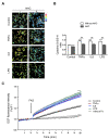
Pro-inflammatory cytokines enhance iron-mediated reactive oxygen species (ROS) production. Hippocampal neurons (7 DIV) were treated with tumor necrosis factor (TNF)α (50 ng/mL), IL6 (50 ng/mL), or lipopolysaccharide (LPS) (1 μg/mL) for 18 h in the presence or absence of 0.5 mM N-acetylcysteine (NAC). (A) The oxidative tone was evaluated by the amount of reduced and oxidized cysteine in proteins. Maleimide-Alexa 488 (green) was used to detect reduced cysteines and maleimide-Alexa 568 (red) to detect oxidized cysteines. The ratio between red and green fluorescence was transformed (ImageJ program) into a thermal scale (right hand bar) in which a shift from blue to red to white implies a higher degree of oxidation. (B) Quantification of the reduced/oxidized cysteine ratio. (C) Increased dichlorofluorescein (DCF) fluorescence, which is a dye sensitive to ROS production, was evaluated after the addition of ferric ammonium sulfate (FAS). Fluorescence data were collected in a microfluorometer plate reader and the ratio between fluorescence (F) and initial fluorescence (Fo) was plotted. Values represent the mean ± SEM (n = 120 neurons, from three independent experiments). *** p < 0.001 compared to the control and ## p < 0.01 compared with the conditions without or with NAC. For protocol detail see [137].
Figure 4
Figure 4
Iron and inflammation are intertwined in a bidirectional relationship during neurodegeneration. The neurodegenerative process starts with the loss of immune homeostatic mechanisms, partially due to decreased norepinephrine (NE) neurotransmission after the degeneration of the locus coeruleus (LC) (1). It continues with neuronal death in intrinsically sensitive areas, such as the substantia nigra (SN) (2a) and the entorhinal cortex (2b), fueled by a positive feedback loop between neuroinflammation, oxidative stress, and iron accumulation (the wheel at the center). As the disease progresses, neurodegeneration continues to other brain regions, such as the hippocampus and the cortex (4). Hepcidin can suppress the main pathologies in experimental Alzheimer’s disease (AD) and Parkinson’s disease (PD) through the inhibition of iron entry at the blood–brain barrier (BBB) (3). Created with BioRender.com.

References

    1. Abe N., Nishihara T., Yorozuya T., Tanaka J. Microglia and Macrophages in the Pathological Central and Peripheral Nervous Systems. Cells. 2020;9:2132. doi: 10.3390/cells9092132. - DOI - PMC - PubMed
    1. Braak H., Ghebremedhin E., Rub U., Bratzke H., Del Tredici K. Stages in the development of Parkinson’s disease-related pathology. Cell Tissue Res. 2004;318:121–134. doi: 10.1007/s00441-004-0956-9. - DOI - PubMed
    1. Sun W., Tang Y., Qiao Y., Ge X., Mather M., Ringman J.M., Shi Y. A probabilistic atlas of locus coeruleus pathways to transentorhinal cortex for connectome imaging in Alzheimer’s disease. NeuroImage. 2020;223:117301. doi: 10.1016/j.neuroimage.2020.117301. - DOI - PMC - PubMed
    1. Braak H., Thal D.R., Ghebremedhin E., Del Tredici K. Stages of the pathologic process in Alzheimer disease: Age categories from 1 to 100 years. J. Neuropathol. Exp. Neurol. 2011;70:960–969. doi: 10.1097/NEN.0b013e318232a379. - DOI - PubMed
    1. Theofilas P., Ehrenberg A.J., Dunlop S., Alho A.T.D.L., Nguy A., Leite R.E.P., Rodriguez R.D., Mejia M.B., Suemoto C.K., Ferretti-Rebustini R.E.L., et al. Locus coeruleus volume and cell population changes during Alzheimer’s disease progression: A stereological study in human postmortem brains with potential implication for early-stage biomarker discovery. Alzheimers Dement. 2017;13:236–246. doi: 10.1016/j.jalz.2016.06.2362. - DOI - PMC - PubMed

LinkOut - more resources