Review
. 2021 Jan 6;13(1):159.
doi: 10.3390/nu13010159.

Regulation of Postabsorptive and Postprandial Glucose Metabolism by Insulin-Dependent and Insulin-Independent Mechanisms: An Integrative Approach

Affiliations
Review

Regulation of Postabsorptive and Postprandial Glucose Metabolism by Insulin-Dependent and Insulin-Independent Mechanisms: An Integrative Approach

George D Dimitriadis et al. Nutrients. .

Abstract

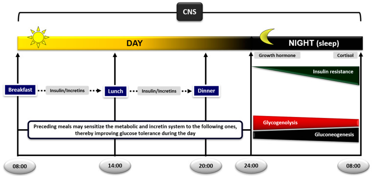
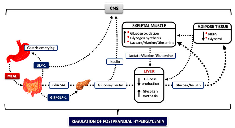
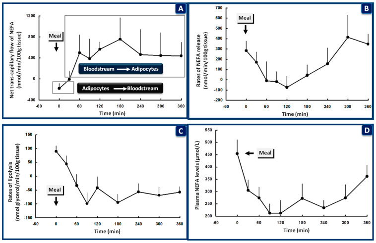
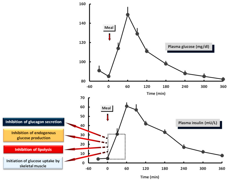
Glucose levels in blood must be constantly maintained within a tight physiological range to sustain anabolism. Insulin regulates glucose homeostasis via its effects on glucose production from the liver and kidneys and glucose disposal in peripheral tissues (mainly skeletal muscle). Blood levels of glucose are regulated simultaneously by insulin-mediated rates of glucose production from the liver (and kidneys) and removal from muscle; adipose tissue is a key partner in this scenario, providing nonesterified fatty acids (NEFA) as an alternative fuel for skeletal muscle and liver when blood glucose levels are depleted. During sleep at night, the gradual development of insulin resistance, due to growth hormone and cortisol surges, ensures that blood glucose levels will be maintained within normal levels by: (a) switching from glucose to NEFA oxidation in muscle; (b) modulating glucose production from the liver/kidneys. After meals, several mechanisms (sequence/composition of meals, gastric emptying/intestinal glucose absorption, gastrointestinal hormones, hyperglycemia mass action effects, insulin/glucagon secretion/action, de novo lipogenesis and glucose disposal) operate in concert for optimal regulation of postprandial glucose fluctuations. The contribution of the liver in postprandial glucose homeostasis is critical. The liver is preferentially used to dispose over 50% of the ingested glucose and restrict the acute increases of glucose and insulin in the bloodstream after meals, thus protecting the circulation and tissues from the adverse effects of marked hyperglycemia and hyperinsulinemia.

Keywords: adipose tissue; fasting; incretins; insulin action secretion; liver; meal sequence; muscle; postabsorptive postprandial glucose metabolism.


Conflict of interest statement

The authors declare no conflict of interest.

Figures

Figure 1
Figure 1
Diurnal regulation of glucose metabolism in the postprandial and postabsorptive state. The sequence of meals plays an important role in postprandial glycemic responses; preceding meals sensitize the metabolic and incretin system to the following ones, thereby improving glucose tolerance during the day. During sleep at night, the gradual development of insulin resistance, due to growth hormone and cortisol surges, ensures that blood glucose levels will be maintained within normal levels until awakening, by switching from glucose to nonesterified fatty acid (NEFA) oxidation in skeletal muscle. The increase in lipolysis and supply of NEFA to the liver and kidneys will also ensure stimulation of gluconeogenesis and glucose production (CNS: central nervous system).
Figure 2
Figure 2
Integrative mechanisms for the regulation of postprandial hyperglycemia. During meal ingestion, several mechanisms operate in concert (gastric emptying and intestinal glucose absorption, secretion and action of gastrointestinal hormones, hyperglycemia mass action effects, and insulin/glucagon secretion and action) to ensure optimal regulation of postprandial glucose fluctuations in blood. Increased concentrations of insulin decrease adipose tissue lipolysis, resulting in a decrease in blood NEFA levels, which then mediate the decrease in endogenous glucose production permitting the initiation of glucose storage, and the increase in glucose uptake by skeletal muscle (CNS: central nervous system; NEFA: nonesterified fatty acids; GIP: glucose-dependent insulinotropic polypeptide; GLP-1: glucagon-like peptide-1).
Figure 3
Figure 3
Nonesterified fatty acid (NEFA) kinetics in the subcutaneous adipose tissue of healthy subjects after a mixed meal given at 08:00, following overnight fasting. At the beginning of the meal (0 time), the trans-capillary flow of NEFA is from the adipocytes to the bloodstream (A). The rates of NEFA release (B), lipolysis (C), and plasma levels of NEFA (D) are all increased to cover energy requirements in muscle during sleep and sustain endogenous glucose production. Within 30 min after the beginning of the meal and in the presence of increasing blood levels of insulin, trans-capillary flow of NEFA is reversed from the bloodstream to the adipocytes (A). The rates of NEFA release (B), lipolysis (C), and plasma levels of NEFA (D) are all rapidly suppressed to allow: (a) storage of the ingested lipids in the adipose tissue; (b) suppression of endogenous glucose production by the liver and kidneys and initiation of glucose storage in the liver; (c) stimulation of glucose uptake in skeletal muscle (data from [149]).
Figure 4
Figure 4
Plasma levels of glucose and insulin after a solid mixed meal in healthy subjects. The early response of insulin: (a) inhibits glucagon secretion from the pancreatic α-cells. (b) Inhibits lipolysis in the adipose tissue. (c) Suppresses endogenous glucose production. (d) Initiates glucose uptake from skeletal muscle. These preliminary effects of insulin start within the first 30 min after the beginning of the meal and are critical for optimal glucose regulation in the postprandial period.

References

    1. Bender D. Introduction to Nutrition and Metabolism. 2nd ed. Taylor & Francis; London, UK: Bristol, PA, USA: 1997. Why eat? pp. 1–10.
    1. Newsholme E.A., Dimitriadis G. Integration of biochemical and physiologic effects of insulin on glucose metabolism. Exp. Clin. Endocrinol. Diabetes. 2001;109:S122–S134. doi: 10.1055/s-2001-18575. - DOI - PubMed
    1. Menting J.G., Whittaker J., Margetts M.B., Whittaker L.J., Kong G.K., Smith B.J., Watson C.J., Zakova L., Kletvikova E., Jiracek J., et al. How insulin engages its primary binding site on the insulin receptor. Nature. 2013;493:241–245. doi: 10.1038/nature11781. - DOI - PMC - PubMed
    1. Kullmann S., Kleinridders A., Small D.M., Fritsche A., Haring H.U., Preissl H., Heni M. Central nervous pathways of insulin action in the control of metabolism and food intake. Lancet Diabetes Endocrinol. 2020;8:524–534. doi: 10.1016/S2213-8587(20)30113-3. - DOI - PubMed
    1. Ward C.W., Lawrence M.C. Landmarks in insulin research. Front. Endocrinol. 2011 doi: 10.3389/fendo.2011.00076. - DOI - PMC - PubMed

LinkOut - more resources