. 2021 Jan 6;11(1):67.
doi: 10.3390/biom11010067.

Diesel Exhaust Particulates Enhances Susceptibility of LPS-Induced Acute Lung Injury through Upregulation of the IL-17 Cytokine-Derived TGF-β1/Collagen I Expression and Activation of NLRP3 Inflammasome Signaling in Mice

Affiliations

Diesel Exhaust Particulates Enhances Susceptibility of LPS-Induced Acute Lung Injury through Upregulation of the IL-17 Cytokine-Derived TGF-β1/Collagen I Expression and Activation of NLRP3 Inflammasome Signaling in Mice

Dong Im Kim et al. Biomolecules. .

Abstract

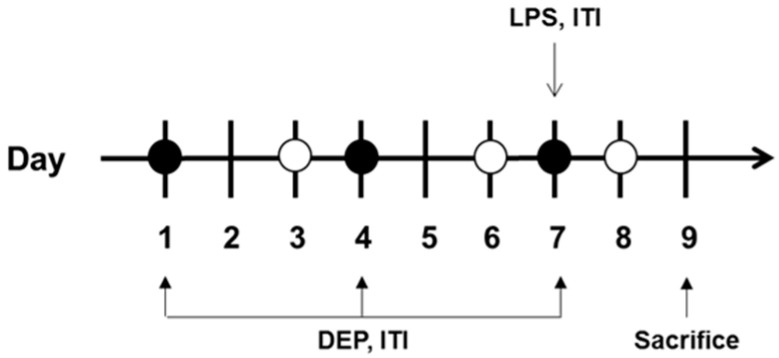
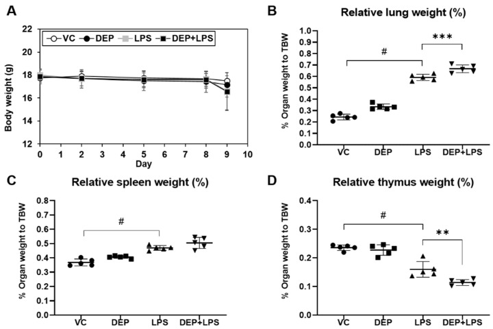
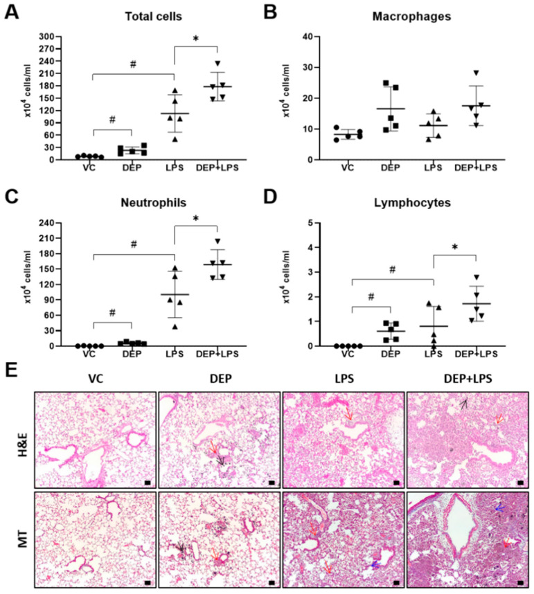
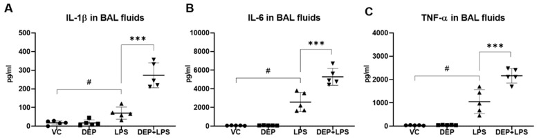
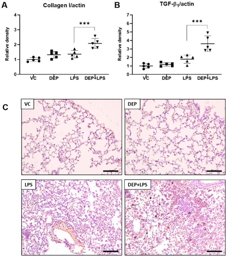
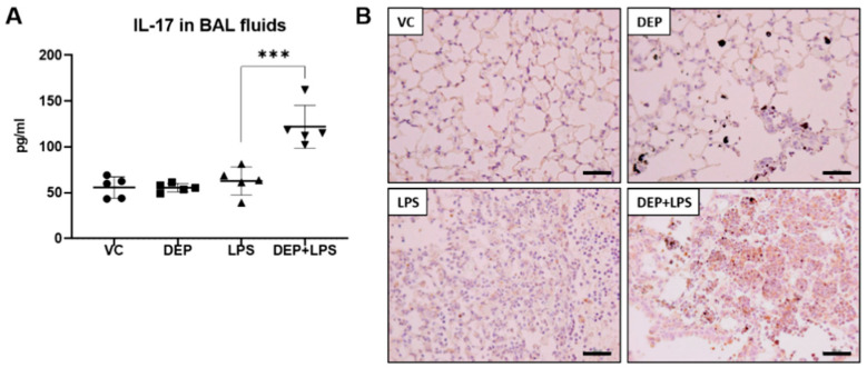
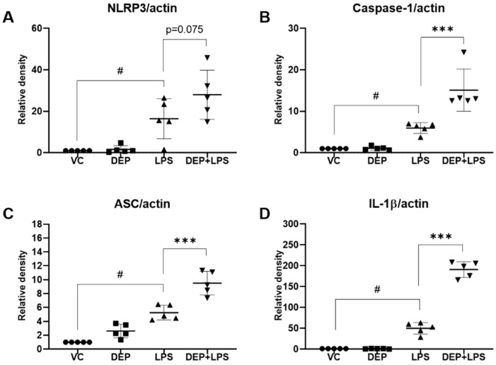
Diesel exhaust particulates (DEP) adversely affect the respiratory system and exacerbate lung diseases, resulting in high mortality rates. However, its pathogenesis is complicated, and the mechanisms involved are incompletely understood. We investigated the effects of DEP pre-exposure on lipopolysaccharide (LPS)-induced acute lung injury (ALI) and identified the roles of interleukin (IL)-17 in mice. Mice were divided into vehicle control, DEP, LPS, and DEP pre-exposed and LPS-instilled groups. Pre-exposure to DEP enhanced the number of total cells, neutrophils, and lymphocytes in the BAL fluid of LPS-instilled mice. Pre-exposure to DEP synergistically exacerbated pulmonary acute lung inflammation and granulomatous inflammation/pulmonary fibrosis, concomitant with the enhanced expression of inflammatory cytokines in the BAL fluid and of collagen I and TGF-β1 in the lungs of LPS-instilled mice. The number of TGF-β1-positive cells in the DEP pre-exposed and LPS-instilled group was higher than that in the LPS group. The expression of NLR family pyrin domain containing 3 (NLRP3) inflammasome components was markedly increased in the DEP pre-exposed and LPS-instilled group. IL-17 levels in the BAL fluid and IL-17-positive cells in the lungs were significantly increased by pre-exposure to DEP in the LPS-induced group compared to that in the DEP or LPS group. These results suggest that DEP predominantly contributes to fibrotic lung disease in LPS-related acute lung injury by upregulating IL-17 cytokine-mediated collagen I and TGF-β1 and, at least in part, by activating LPS-induced NLRP3 inflammasome signaling. The study should be useful in devising better strategies for prevention and management of ALI.

Keywords: IL-17; NLRP3; TGF-β1; acute lung injury; diesel exhaust particulate; lung fibrosis.


Conflict of interest statement

The authors declare no conflict of interest.

Figures

Figure 1
Figure 1
Diagram showing the in vivo experimental protocol. DEP, diesel exhaust particulate; LPS, lipopolysacharide; ITI, intratracheal injection.
Figure 2
Figure 2
Changes in (A) body weight and (BD) relative organ weight in vehicle control (VC), DEP, LPS, DEP pre-exposed, and LPS-instilled (DEP+LPS) groups. Relative weights of the (B) lung, (C) spleen, and (D) thymus were calculated using the following formula: relative organ weight = organ weight (g)/final body weight (g) × 100%. Data represent means ± SD (n = 5 per group). # p < 0.05 vs. VC, ** p < 0.01 or *** p < 0.001 vs. LPS group.
Figure 3
Figure 3
(AD) Cellular changes in the bronchoalveolar lavage (BAL) fluid and (E) representative hematoxylin and eosin (H&E) and Masson’s trichrome-stained images of the lung sections from the vehicle control (VC), DEP, LPS, and DEP pre-exposed and LPS-instilled (DEP + LPS) groups. Black, red, and blue arrows indicate particle-pigmented alveolar macrophages, inflammatory infiltration, and collagen deposition, respectively. Scale bars = 50 μm. Data represent means ± SD (n = 5 per group). # p < 0.05 vs. VC, * p < 0.05 vs. LPS group.
Figure 4
Figure 4
Protein levels of (A) IL-1β, (B) IL-6, and (C) TNF-α in the bronchoalveolar (BAL) fluid from mice in the vehicle control (VC), DEP, LPS, and DEP pre-exposed and LPS-instilled (DEP+LPS) groups. Data represent means ± SD (n = 5 per group). # p < 0.05 vs. VC, *** p < 0.001 vs. LPS group.
Figure 5
Figure 5
Protein levels of (A) collagen I and (B) TGF-β1 and representative images of immunohistochemistry for TGF-β1 ((C), brown color) in the lung tissue sections from mice in the VC, DEP, LPS, and DEP pre-exposed and LPS-instilled (DEP+LPS) groups. Scale bars = 500 nm. Data represent means ± SD (n = 5 per group). *** p < 0.001 vs. LPS group.
Figure 6
Figure 6
Protein levels of (A) IL-17 in the bronchoalveolar (BAL) fluid and (B) representative images of immunohistochemistry (IHC) for IL-17 (brown color) in the lung tissue sections from mice in the VC, DEP, LPS, and DEP pre-exposed and LPS-instilled (DEP+LPS) groups. Scale bars = 200 nm. Data represent means ± SD (n = 5 per group) *** p < 0.001 vs. LPS group.
Figure 7
Figure 7
Protein levels of (A) NLRP3, (B) caspase-1, (C) ASC, and (D) IL-1β in the lung tissue of mice in the vehicle control (VC), DEP, LPS, and DEP pre-exposed and LPS-instilled (DEP+LPS) groups. Data represent means ± SD (n = 5 per group). # p < 0.05 vs. VC, *** p < 0.001 vs. LPS group.

Similar articles

Cited by

References

    1. Matthay M.A., Zemans R.L., Zimmerman G.A., Arabi Y.M., Beitler J.R., Mercat A., Herridge M., Randolph A.G., Calfee C.S. Acute respiratory distress syndrome. Nat. Rev. Dis. Primers. 2019;5:18. doi: 10.1038/s41572-019-0069-0. - DOI - PMC - PubMed
    1. Moazed F., Calfee C.S. Environmental risk factors for ARDS. Clin. Chest Med. 2014;35:625–637. doi: 10.1016/j.ccm.2014.08.003. - DOI - PMC - PubMed
    1. Strosnider H.M., Chang H.H., Darrow L.A., Liu Y., Vaidyanathan A., Strickland M.J. Age-specific associations of ozone and fine particulate matter with respiratory emergency department visits in the United States. Am. J. Respir. Crit. Care Med. 2019;199:882. doi: 10.1164/rccm.201806-1147OC. - DOI - PubMed
    1. Schulze F., Gao X., Virzonis D., Damiati S., Schneider M.R., Kodzius R. Air quality effects on human health and approaches for its assessment through microfluidic chips. Genes. 2017;8:244. doi: 10.3390/genes8100244. - DOI - PMC - PubMed
    1. Lin H., Tao J., Kan H., Qian Z., Chen A., Du Y., Liu T., Zhang Y., Qi Y., Ye J., et al. Ambient particulate matter air pollution associated with acute respiratory distress syndrome in Guangzhou, China. J. Expo. Sci. Environ. Epidemiol. 2018;28:392–399. doi: 10.1038/s41370-018-0034-0. - DOI - PubMed

Publication types

MeSH terms

LinkOut - more resources