Cryptococcus in Wildlife and Free-Living Mammals
- PMID: 33419125
- PMCID: PMC7825559
- DOI: 10.3390/jof7010029
Cryptococcus in Wildlife and Free-Living Mammals
Abstract
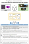
Cryptococcosis is typically a sporadic disease that affects a broad range of animal species globally. Disease is a consequence of infection with members of the Cryptococcus neoformans or Cryptococcus gattii species complexes. Although cryptococcosis in many domestic animals has been relatively well-characterized, free-living wildlife animal species are often neglected in the literature outside of occasional case reports. This review summarizes the clinical presentation, pathological findings and potential underlying causes of cryptococcosis in various other animals, including terrestrial wildlife species and marine mammals. The evaluation of the available literature supports the hypothesis that anatomy (particularly of the respiratory tract), behavior and environmental exposures of animals play vital roles in the outcome of host-pathogen-environment interactions resulting in different clinical scenarios. Key examples range from koalas, which exhibit primarily C. gattii species complex disease presumably due to their behavior and environmental exposure to eucalypts, to cetaceans, which show predominantly pulmonary lesions due to their unique respiratory anatomy. Understanding the factors at play in each clinical scenario is a powerful investigative tool, as wildlife species may act as disease sentinels.
Keywords: C. gattii; C. neoformans; Cryptococcus; cryptococcosis; felid; koala; marine; wildlife.
Conflict of interest statement
The authors declare no conflict of interest.
Figures


Similar articles
-
Jet-Setting Koalas Spread Cryptococcus gattii VGII in Australia.mSphere. 2019 Jun 5;4(3):e00216-19. doi: 10.1128/mSphere.00216-19. mSphere. 2019. PMID: 31167945 Free PMC article.
-
Prevalence of cryptococcal antigenemia and nasal colonization in a free-ranging koala population.Med Mycol. 2019 Oct 1;57(7):848-857. doi: 10.1093/mmy/myy144. Med Mycol. 2019. PMID: 30649397
-
Long-term surveillance and treatment of subclinical cryptococcosis and nasal colonization by Cryptococcus neoformans and C. gattii species complex in captive koalas (Phascolarctes cinereus).Med Mycol. 2012 Apr;50(3):291-8. doi: 10.3109/13693786.2011.594967. Epub 2011 Aug 23. Med Mycol. 2012. PMID: 21859391
-
[Cryptococcosis caused by Cryptococcus neoformans var. Gattii. A case associated with acquired immunodeficiency syndrome (AIDS) in Kinshasa, Zaire].Med Trop (Mars). 1992 Oct-Dec;52(4):435-8. Med Trop (Mars). 1992. PMID: 1494313 Review. French.
-
Equine Pulmonary Cryptococcosis: A Comparative Literature Review and Evaluation of Fluconazole Monotherapy.Mycopathologia. 2017 Apr;182(3-4):413-423. doi: 10.1007/s11046-016-0065-9. Epub 2016 Sep 21. Mycopathologia. 2017. PMID: 27655152 Review.
Cited by
-
Genetic diversity and antifungal susceptibilities of environmental Cryptococcus neoformans and Cryptococcus gattii species complexes.IMA Fungus. 2024 Jul 25;15(1):21. doi: 10.1186/s43008-024-00153-w. IMA Fungus. 2024. PMID: 39060926 Free PMC article.
-
First case of feline cryptococcosis in Bosnia and Herzegovina.JFMS Open Rep. 2024 Aug 7;10(2):20551169241265248. doi: 10.1177/20551169241265248. eCollection 2024 Jul-Dec. JFMS Open Rep. 2024. PMID: 39131486 Free PMC article.
-
An analysis of the population of Cryptococcus neoformans strains isolated from animals in Poland, in the years 2015-2019.Sci Rep. 2021 Mar 23;11(1):6639. doi: 10.1038/s41598-021-86169-3. Sci Rep. 2021. PMID: 33758319 Free PMC article.
-
Pulmonary Cryptococcosis.J Fungi (Basel). 2022 Oct 31;8(11):1156. doi: 10.3390/jof8111156. J Fungi (Basel). 2022. PMID: 36354923 Free PMC article. Review.
-
A Possible Link between the Environment and Cryptococcus gattii Nasal Colonisation in Koalas (Phascolarctos cinereus) in the Liverpool Plains, New South Wales.Int J Environ Res Public Health. 2022 Apr 11;19(8):4603. doi: 10.3390/ijerph19084603. Int J Environ Res Public Health. 2022. PMID: 35457470 Free PMC article.
References
-
- Kwon-Chung K.J., Bennett J.E., Wickes B.L., Meyer W., Cuomo C.A., Wollenburg K.R., Bicanic T.A., Castañeda E., Chang Y.C., Chen J., et al. The Case for Adopting the “Species Complex” Nomenclature for the Etiologic Agents of Cryptococcosis. mSphere. 2017;2:1–7. doi: 10.1128/mSphere.00357-16. - DOI - PMC - PubMed
Publication types
LinkOut - more resources
Full Text Sources
Other Literature Sources

