

Ferroptosis-Related Genes in Neurodevelopment and Central Nervous System
- PMID: 33419148
- PMCID: PMC7825574
- DOI: 10.3390/biology10010035
Ferroptosis-Related Genes in Neurodevelopment and Central Nervous System
Abstract
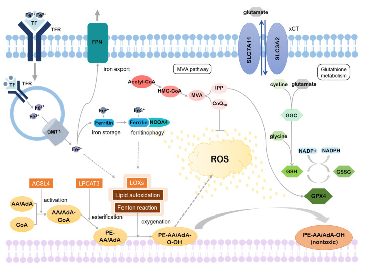
Ferroptosis, first introduced as a new form of regulated cell death induced by erastin, is accompanied by the accumulation of iron and lipid peroxides, thus it can be inhibited either by iron chelators or by lipophilic antioxidants. In the past decade, multiple studies have introduced the potential importance of ferroptosis in many human diseases, including cancer and neurodegenerative diseases. In this review, we will discuss the genetic association of ferroptosis with neurological disorders and development of the central nervous system.
Keywords: CNS; brain development; ferroptosis; iron; lipid peroxides; neurological disorder.
Conflict of interest statement
The authors declare no conflict of interest.
Figures
Similar articles
-
Mechanisms of Ferroptosis and Relations With Regulated Cell Death: A Review.Front Physiol. 2019 Feb 26;10:139. doi: 10.3389/fphys.2019.00139. eCollection 2019. Front Physiol. 2019. PMID: 30863316 Free PMC article. Review.
-
Role of ferroptosis in neurological diseases.Neurosci Lett. 2021 Mar 16;747:135614. doi: 10.1016/j.neulet.2020.135614. Epub 2021 Jan 21. Neurosci Lett. 2021. PMID: 33485988 Review.
-
Alpha-synuclein interaction with mitochondria is the final mechanism of ferroptotic death induced by erastin in SH-SY5Y cells.Free Radic Res. 2024 Mar;58(3):217-228. doi: 10.1080/10715762.2024.2336563. Epub 2024 Apr 5. Free Radic Res. 2024. PMID: 38572725
-
Dysregulation of Iron Homeostasis in the Central Nervous System and the Role of Ferroptosis in Neurodegenerative Disorders.Antioxid Redox Signal. 2022 Jul;37(1-3):150-170. doi: 10.1089/ars.2021.0218. Epub 2022 Jan 4. Antioxid Redox Signal. 2022. PMID: 34569265 Review.
-
Curcumin and (-)- Epigallocatechin-3-Gallate Protect Murine MIN6 Pancreatic Beta-Cells Against Iron Toxicity and Erastin-Induced Ferroptosis.Pharmaceuticals (Basel). 2019 Feb 6;12(1):26. doi: 10.3390/ph12010026. Pharmaceuticals (Basel). 2019. PMID: 30736288 Free PMC article.
Cited by
-
Ferroptosis contributes to hypoxic-ischemic brain injury in neonatal rats: Role of the SIRT1/Nrf2/GPx4 signaling pathway.CNS Neurosci Ther. 2022 Dec;28(12):2268-2280. doi: 10.1111/cns.13973. Epub 2022 Oct 2. CNS Neurosci Ther. 2022. PMID: 36184790 Free PMC article.
-
Identification of the Ferroptosis-Associated Gene Signature to Predict the Prognostic Status of Endometrial Carcinoma Patients.Comput Math Methods Med. 2021 Sep 7;2021:9954370. doi: 10.1155/2021/9954370. eCollection 2021. Comput Math Methods Med. 2021. PMID: 34531924 Free PMC article.
-
An Original Ferroptosis-Related Gene Signature Effectively Predicts the Prognosis and Clinical Status for Colorectal Cancer Patients.Front Oncol. 2021 Jun 24;11:711776. doi: 10.3389/fonc.2021.711776. eCollection 2021. Front Oncol. 2021. PMID: 34249766 Free PMC article.
-
A Comprehensive Study of De Novo Mutations on the Protein-Protein Interaction Interfaces Provides New Insights into Developmental Delay.Biomolecules. 2022 Nov 6;12(11):1643. doi: 10.3390/biom12111643. Biomolecules. 2022. PMID: 36358993 Free PMC article.
-
Exploring the key ferroptosis-related gene in the peripheral blood of patients with Alzheimer's disease and its clinical significance.Front Aging Neurosci. 2022 Sep 1;14:970796. doi: 10.3389/fnagi.2022.970796. eCollection 2022. Front Aging Neurosci. 2022. PMID: 36118694 Free PMC article.
References
Publication types
Grants and funding
LinkOut - more resources
Full Text Sources
Other Literature Sources
Miscellaneous
