. 2020 Dec 20;9(12):2720.
doi: 10.3390/cells9122720.

Implication of the Association of Fibrinogen Citrullination and Osteoclastogenesis in Bone Destruction in Rheumatoid Arthritis

Affiliations

Implication of the Association of Fibrinogen Citrullination and Osteoclastogenesis in Bone Destruction in Rheumatoid Arthritis

Ji Soo Kim et al. Cells. .

Abstract

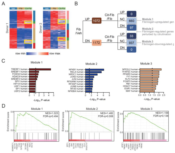
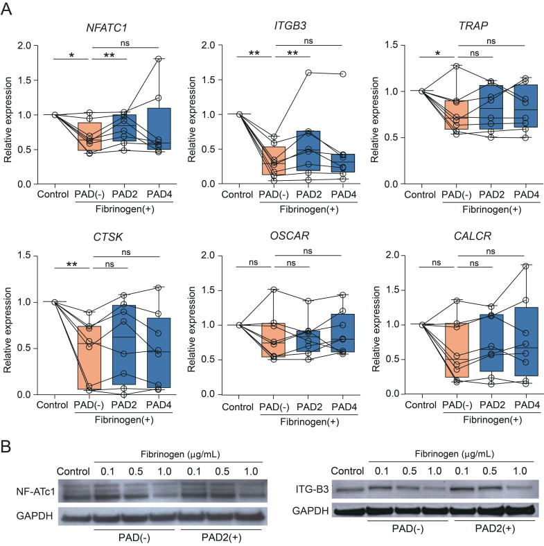
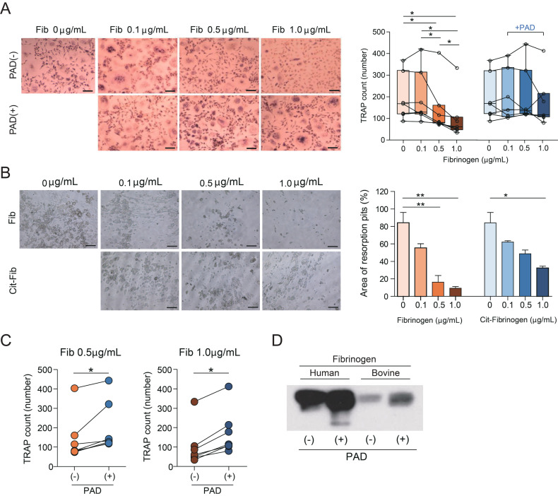
Immune complexes containing citrullinated fibrinogen are present in the sera and synovium of rheumatoid arthritis patients and potentially contribute to synovitis. However, fibrinogen can inhibit the osteoclastogenesis of precursor cells. We investigated the direct effect of citrullinated fibrinogen on osteoclastogenesis to understand the role of citrullination on bone erosion of rheumatoid arthritis patients. We evaluated the fibrinogen citrullination sites using mass spectrometry and quantified osteoclast-related protein and gene expression levels by Western blotting, microarray, and real-time polymerase chain reaction. Differences in spectral peaks were noted between fibrinogen and citrullinated fibrinogen at five sites in α-chains, two sites in β-chains, and one site in a γ-chain. Transcriptome changes induced by fibrinogen and citrullinated fibrinogen were identified and differentially expressed genes grouped into three distinctive modules. Fibrinogen was then citrullinated in vitro using peptidylarginine deiminase. When increasing doses of soluble fibrinogen and citrullinated fibrinogen were applied to human CD14+ monocytes, citrullination restored osteoclastogenesis-associated changes, including NF-ATc1 and ß3-integrin. Finally, citrullination rescued the number of osteoclasts by restoring fibrinogen-induced suppression of osteoclastogenesis. Taken together, the results indicate that the inhibitory function of fibrinogen on osteoclastogenesis is reversed by citrullination and suggest that citrullinated fibrinogen may contribute to erosive bone destruction in rheumatoid arthritis.

Keywords: citrullinated fibrinogen; osteoclastogenesis; rheumatoid arthritis.


Conflict of interest statement

The authors declare no conflict of interest.

Figures

Figure 1
Figure 1
Citrullination of fibrinogen at α, β, and γ chains in the presence of peptidylarginine deiminase (PAD). (A) Level of citrullinated fibrinogen in synovial fluid (SF) from two osteoarthritis (OA SF1, OA SF2) and two rheumatoid arthritis (RA SF1, RA SF2) patients was detected by immunoprecipitation (IP) with anti-fibrinogen antibody and Western blot (WB) using anti-modified citrulline antibody. Coomassie, corresponding to each WB, of total protein in synovial fluid. Arrows indicate corresponding bands for α, β, and γ chains of fibrinogen. Fibrinogen from RA SF was more citrullinated than OA SF. (B,C) Spots for the α, β, and γ chains shifted in two-dimensional mapping of fibrinogen in the absence (B, upper row) or presence (C, upper row) of PAD2 (PAD). Shifts due to citrullination were confirmed by WB using anti-citrullinated fibrinogen antibody (lower rows of B and C). Arrows indicate corresponding spots between two-dimensional electrophoresis (2DE) and WB at a designated pH. (D) Citrullination sites (five in α, two in β, and one in γ chains) were detected by mass spectrometry via modification with phenylglyoxal monohydrate (PGM). Top panel = bovine fibrinogen (Fib.), Middle panel = Fib. + PAD, Bottom panel = Fib. + PAD + PGM.
Figure 2
Figure 2
Differentially expressed genes (DEGs) in CD14+ sorted monocytes from two peripheral blood donors in the presence of fibrinogen or citrullinated fibrinogen. (A) Heatmap of DEGs. Veh = vehicle, Fib = bovine fibrinogen, Cit-Fib = bovine fibrinogen citrullinated by PAD2. Six different clusters of DEGs were presented by K-means clustering analysis. Yellow boxes indicate the clusters associated with restoration of fibrinogen-induced change by citrullination. (B) Fibrinogen-regulated genes perturbed by citrullination correspond to DEGs in the yellow box in (A). UP = upregulated genes, NC = no change in expression, DN = downregulated genes. (C) Gene set enrichment analysis of the module 1, 2, or 3 using the human TRRUST transcription factor database. Dashed line = p-value 0.05. (D) Gene set enrichment analysis of the module 1, 2, or 3 using osteoclast-specific gene set originated from ref. 21. NES = normalized enrichment score, FDR-q = q-value of false discovery rate.
Figure 3
Figure 3
Gene and protein expression in osteoclasts in the presence of PAD. (A) Real-time PCR of cells cultured in the absence or presence of PAD2 or PAD4 at a dose of 1.0 μg/mL (n = 8). * p  <  0.05; ** p  <  0.01; ns—not significant. Individual values are plotted, and error bars represent SD. (B) Western blot of NF-ATc1, β3-integrin, and GAPDH. M-CSF and RANKL were supplemented in the control group.
Figure 4
Figure 4
Reversion of dose-dependent inhibition of osteoclastogenesis in the presence of PAD. (A) Left: monocytes were cultured in the absence or presence of PAD2 (PAD) and stained for TRAP on day 6 (n = 6). M-CSF (20 ng/mL) and RANKL (40 ng/mL) were replenished every 3 days. M-CSF and RANKL were supplemented in the control group. Fib = bovine fibrinogen. Scale bar 200 μm. Right: number of TRAP-positive osteoclasts in the absence or presence of PAD. * p  <  0.05. Individual values are plotted, and error bars represent SD. (B) Left: bone resorption activity in the presence of fibrinogen or citrullinated fibrinogen for 6 days. Fib = human fibrinogen, Cit-Fib = citrullinated human fibrinogen. Scale bar 100 μm. Right: area of resorption pits (%). * p  <  0.05; ** p  <  0.01. (C) Number of TRAP-positive osteoclasts according to fibrinogen dose. * p  <  0.05. Individual values are plotted. (D) Western blot using an antibody against citrullinated fibrinogen and protein from reaction mixtures containing PAD with human fibrinogen or bovine fibrinogen incubated at 37 °C for 2 h.

Similar articles

Cited by

References

    1. Doolittle R.F., Spraggon G., Everse S.J. Three-dimensional structural studies on fragments of fibrinogen and fibrin. Curr. Opin. Struct. Biol. 1998;8:792–798. doi: 10.1016/S0959-440X(98)80100-0. - DOI - PubMed
    1. Herrick S., Blanc-Brude O., Gray A., Laurent G. Fibrinogen. Int. J. Biochem. Cell Biol. 1999;31:741–746. doi: 10.1016/S1357-2725(99)00032-1. - DOI - PubMed
    1. Haidaris P.J., Francis C.W., Sporn L.A., Arvan D.S., Collichio F.A., Marder V.J. Megakaryocyte and hepatocyte origins of human fibrinogen biosynthesis exhibit hepatocyte-specific expression of gamma chain-variant polypeptides. Blood. 1989;74:743–750. doi: 10.1182/blood.V74.2.743.743. - DOI - PubMed
    1. Adams R.A., Schachtrup C., Davalos D., Tsigelny I., Akassoglou K. Fibrinogen signal transduction as a mediator and therapeutic target in inflammation: Lessons from multiple sclerosis. Curr. Med. Chem. 2007;14:2925–2936. doi: 10.2174/092986707782360015. - DOI - PubMed
    1. Lishko V.K., Kudryk B., Yakubenko V.P., Yee V.C., Ugarova T.P. Regulated unmasking of the cryptic binding site for integrin alpha M beta 2 in the gamma C-domain of fibrinogen. Biochemistry. 2002;41:12942–12951. doi: 10.1021/bi026324c. - DOI - PubMed

Publication types

MeSH terms